File:Patten005.jpg
Original file (614 × 868 pixels, file size: 137 KB, MIME type: image/jpeg)
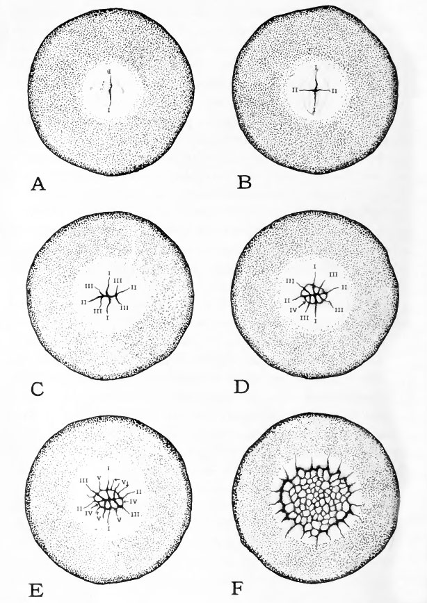
Fig. 5. Surface aspect of blastoderm at various stages of cleavage
(Based on Blount's photomicrographs of the pigeon's egg.) The blastoderm and the immediately surrounding yolk are viewed directly from the animal pole, the shell and albumen having been removed.
The order in which vthe cleavage furrows have appeared is indicated on the diagrams by Roman numerals.
A, first cleavage; B, second cleavage
C, third cleavage; D, fourth cleavage
E, fifth cleavage; F, early morula.
- Links: Introduction | Gametes and Fertilization | Segmentation | Entoderm | Primitive Streak and Mesoderm | Primitive Streak to Somites | 24 Hours | 24 to 33 Hours | 33 to 39 Hours | 40 to 50 Hours | Extra-embryonic Membranes | 50 to 55 Hours | Day 3 to 4 | References | All Figures
| Historic Disclaimer - information about historic embryology pages |
|---|
 |
Reference
Patten BM. The Early Embryology of the Chick. (1920) Philadelphia: P. Blakiston's Son and Co.
Cite this page: Hill, M.A. (2024, April 25) Embryology Patten005.jpg. Retrieved from https://embryology.med.unsw.edu.au/embryology/index.php/File:Patten005.jpg
- © Dr Mark Hill 2024, UNSW Embryology ISBN: 978 0 7334 2609 4 - UNSW CRICOS Provider Code No. 00098G
File history
Click on a date/time to view the file as it appeared at that time.
| Date/Time | Thumbnail | Dimensions | User | Comment | |
|---|---|---|---|---|---|
| current | 15:59, 14 January 2011 | | 614 × 868 (137 KB) | S8600021 (talk | contribs) | ==Fig. 5. Surface aspect of blastoderm at various stages of cleavage== (Based on Blount's photomicrographs of the pigeon's egg.) The blastoderm and the immediately surrounding yolk are viewed directly from the animal pole, the shell and albumen having be |
You cannot overwrite this file.
File usage
The following 2 pages use this file: