File:Ovary histology 004.jpg
Original file (1,280 × 1,024 pixels, file size: 401 KB, MIME type: image/jpeg)
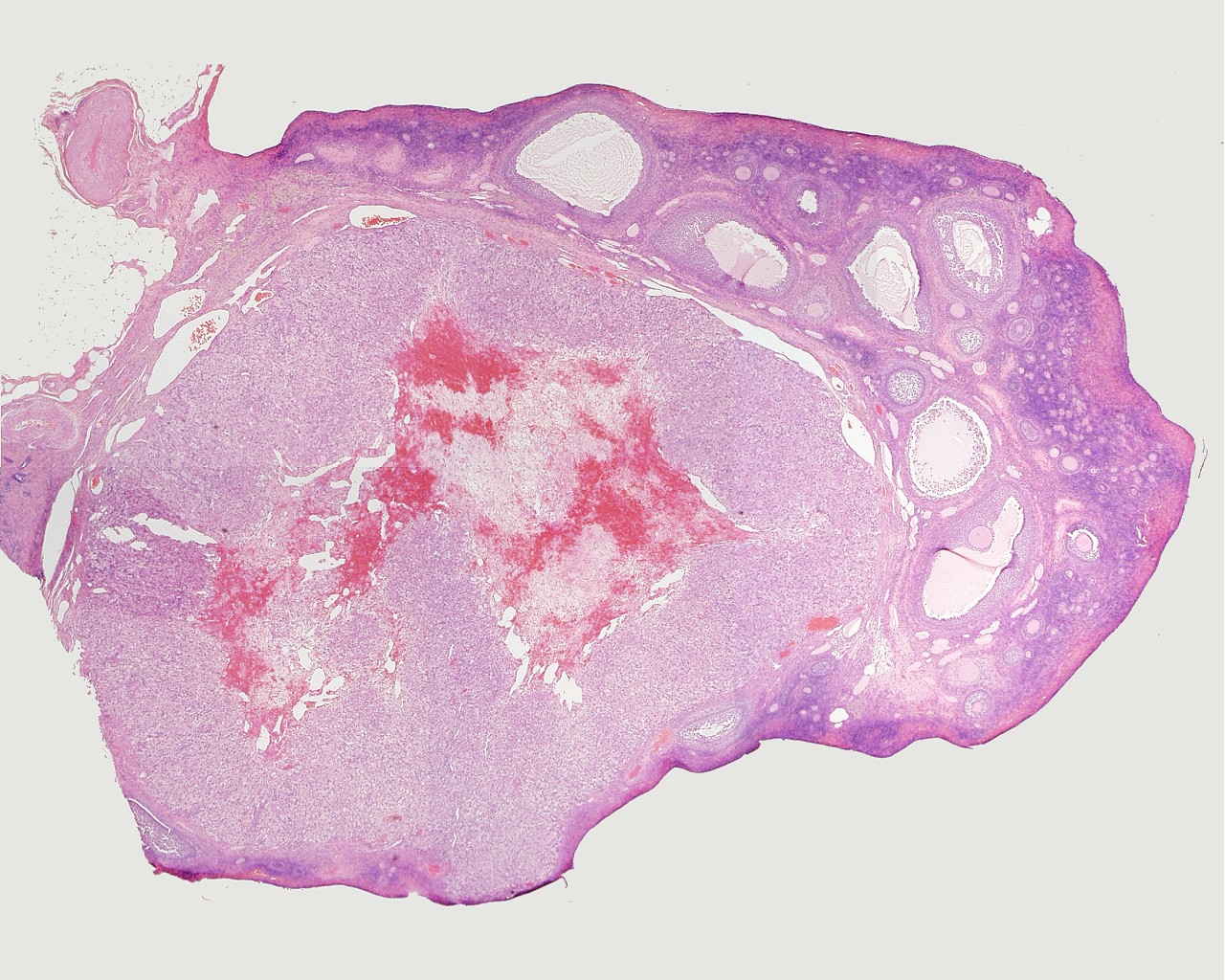
Ovary - Corpus Luteum
Histology image shows the corpus luteum (Latin, corpus = body; luteum = yellow)
- The ovulating follicle forms initially the corpus hemorrhagicum and then corpus luteum.
- Corpus luteum (CL, yellow body) outer layer consisting of theca lutein cells and more centrally the granulosa lutein cells.
- Corpus luteum core is initially filled with a blood clot, following ovulation, and is the invaded by connective tissue.
Theca lutein cells and granulosa lutein cells work together in the production of ovarian hormones that support the initial pregnancy. Lack of implantation and associated hCG will lead to this structure not producing hormones and forming the corpus albicans (white body).
Hormone secretion in the corpus luteum ceases within 14 days after ovulation if the oocyte is not fertilised. In this case, the corpus luteum degenerates into a corpus albicans - whitish scar tissue within the ovaries.
Hormone secretion continues for 2-3 month after ovulation if fertilisation occurs.
The dark cell represent theca lutein cell the lighter ones are granulosa lutein cells.
File:Ovary_histology_004.jpg
Ovary histology: Tunica Albuginea x20 | Tunica albuginea, Germinal epithelium x40 |
Primary follicle, primordial follicle, oocyte, x40 | Secondary follicle, cumulus oophorus, zona pelucida, granulosa cells, oocyte x20 | Corpus luteum, theca lutein cells, granulosa lutein cells, Loupe | Corpus luteum, theca lutein cells, granulosa lutein cells, x10 | Corpus luteum, theca lutein cells, granulosa lutein cells, x40 | Corpus albicans, primary follicle, primordial follicle, granulosa cells, oocyte x20 | Menstrual Cycle | Ovary Development
Links: Histology | Histology Stains | Blue Histology images copyright Lutz Slomianka 1998-2009. The literary and artistic works on the original Blue Histology website may be reproduced, adapted, published and distributed for non-commercial purposes. See also the page Histology Stains.
Cite this page: Hill, M.A. (2024, April 23) Embryology Ovary histology 004.jpg. Retrieved from https://embryology.med.unsw.edu.au/embryology/index.php/File:Ovary_histology_004.jpg
- © Dr Mark Hill 2024, UNSW Embryology ISBN: 978 0 7334 2609 4 - UNSW CRICOS Provider Code No. 00098G
Ovary, monkey H&E reproductive system, female, overview, corpus luteum Loupe
File history
Click on a date/time to view the file as it appeared at that time.
| Date/Time | Thumbnail | Dimensions | User | Comment | |
|---|---|---|---|---|---|
| current | 21:09, 23 February 2011 | | 1,280 × 1,024 (401 KB) | S8600021 (talk | contribs) | File:Ovary_histology_004.jpg Ovary,_monkey_H&E_reproductive_system,_female,_overview,_corpus_luteum_Loupe.jpg {{Ovary Histology}} {{Template:Blue Histology}} Category:Monkey Category:Genital Category:Histology Category:Ovary [[Catego |
You cannot overwrite this file.
File usage
The following 5 pages use this file: