File:Ova41he.jpg
Ova41he.jpg (450 × 600 pixels, file size: 113 KB, MIME type: image/jpeg)
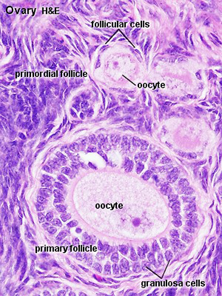
Ovary - Primary Follicle
Histological image showing both primordial follicles and a single primary follicle.
There are several different nomenclatures for the stages of follicle maturation. It probably does not matter which naming system you use, as long as you are consistent and use the same set of terminology for all stages. Early stages of follicle development appear to be gonadotropin (Gn) independent and with development become gonadotropin "sensitive" and then "dependent" . (UK spelling is gonadotrophin).
- Primordial Follicle - Alternate nomenclature: small follicle or type 1, 2, 3 (25cells)
- Primary Follicle - Alternate nomenclature: preantral follicle or type 4 (26-100 cells), type 5 (101-300 cells)
- Secondary Follicle - Alternate nomenclature: small and large antral follicle or type 6 (3001-500 cells), type 7 (501-1000 cells)
- Preovulatory Follicle - Alternate nomenclature: Graafian follicle or type 8 (>1000 cells)
- Atresia - At any one time the majority of follicles are destined not to complete maturation and at any stage (from type 4-7) degeneration of the follicle can occur.
- Links: Oocyte Development | Ovary Development
Ovary histology: Tunica Albuginea x20 | Tunica albuginea, Germinal epithelium x40 | Primary follicle, primordial follicle, oocyte, x40 | Secondary follicle, cumulus oophorus, zona pelucida, granulosa cells, oocyte x20 | Corpus luteum, theca lutein cells, granulosa lutein cells, Loupe | Corpus luteum, theca lutein cells, granulosa lutein cells, x10 | Corpus luteum, theca lutein cells, granulosa lutein cells, x40 | Corpus albicans, primary follicle, primordial follicle, granulosa cells, oocyte x20 | Menstrual Cycle | Ovary Development
Histology image H&E high power
Links: Histology | Histology Stains | Blue Histology images copyright Lutz Slomianka 1998-2009. The literary and artistic works on the original Blue Histology website may be reproduced, adapted, published and distributed for non-commercial purposes. See also the page Histology Stains.
Cite this page: Hill, M.A. (2024, April 19) Embryology Ova41he.jpg. Retrieved from https://embryology.med.unsw.edu.au/embryology/index.php/File:Ova41he.jpg
- © Dr Mark Hill 2024, UNSW Embryology ISBN: 978 0 7334 2609 4 - UNSW CRICOS Provider Code No. 00098G
Ova41he.jpg
File history
Click on a date/time to view the file as it appeared at that time.
| Date/Time | Thumbnail | Dimensions | User | Comment | |
|---|---|---|---|---|---|
| current | 16:32, 6 May 2012 | | 450 × 600 (113 KB) | Z8600021 (talk | contribs) | |
| 11:17, 28 July 2009 |  | 300 × 400 (66 KB) | MarkHill (talk | contribs) | Ovary Histology Source: UWA Blue Histology |
You cannot overwrite this file.
File usage
The following 20 pages use this file:
- 2009 Lecture 2
- 2010 BGD Lecture - Development of the Embryo/Fetus 1
- 2010 BGD Practical 3 - Oogenesis and Ovulation
- 2010 BGD Practical 3 - Week 3 Summary
- 2010 Lab 2
- 2010 Lecture 2
- 2011 Lab 1 - Oogenesis
- ANAT2341 Lab 1 - Oogenesis
- BGDA Lecture - Development of the Embryo/Fetus 1
- BGDA Lecture - Development of the Embryo/Fetus 2
- BGDA Practical 3 - Oogenesis and Ovulation
- BGDA Practical 3 - Week 3 Summary
- Fertilization
- Histology Stains
- Lecture - Fertilization
- Ovary Development
- Timeline human development
- Talk:Timeline human development
- Template:First Trimester Timeline
- Template:First Trimester Timeline collapsable table