File:OtisBrent1954 fig01.jpg
From Embryology
Size of this preview: 650 × 599 pixels. Other resolution: 1,280 × 1,180 pixels.
Original file (1,280 × 1,180 pixels, file size: 144 KB, MIME type: image/jpeg)
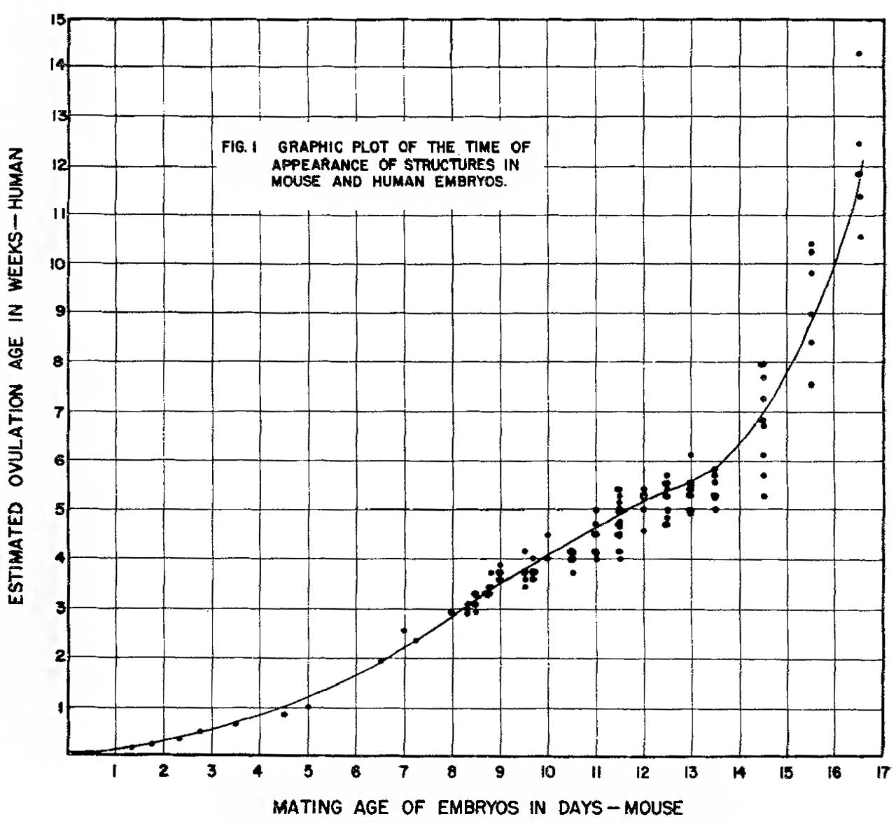
Fig. 1. Graphic plot of the times of appearance of structures in mouse and human embryos
| Historic Disclaimer - information about historic embryology pages |
|---|
 |
Mouse and human embryos: plate 1 | plate 2 | plate 3 | plate 4 | 1954 Otis Brent | mouse
Reference
Otis EM and Brent R. Equivalent ages in mouse and human embryos. (1954) Anat Rec. 120(1):33-63. PMID 13207763
Cite this page: Hill, M.A. (2024, April 16) Embryology OtisBrent1954 fig01.jpg. Retrieved from https://embryology.med.unsw.edu.au/embryology/index.php/File:OtisBrent1954_fig01.jpg
- © Dr Mark Hill 2024, UNSW Embryology ISBN: 978 0 7334 2609 4 - UNSW CRICOS Provider Code No. 00098G
File history
Click on a date/time to view the file as it appeared at that time.
| Date/Time | Thumbnail | Dimensions | User | Comment | |
|---|---|---|---|---|---|
| current | 13:31, 31 May 2018 | | 1,280 × 1,180 (144 KB) | Z8600021 (talk | contribs) | ===Reference=== {{Ref-OtisBrent1954}} {{Footer}} |
You cannot overwrite this file.
File usage
The following 2 pages use this file: