File:Otic placode embryo.jpg
Otic_placode_embryo.jpg (500 × 461 pixels, file size: 23 KB, MIME type: image/jpeg)
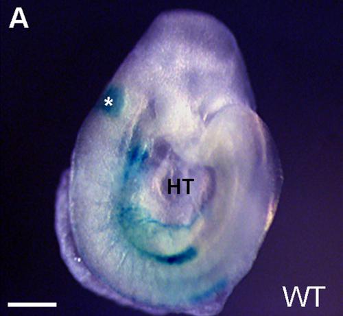
Mouse Otic Placode
The lateral view of an E8.5 embryo used as the control Hoxa3-lacZ (normal wild-type). Asterisk indicates the otic vesicle. HT, heart tube. Scale bars = 100 µm.
Reference
<pubmed>22110697</pubmed>
Copyright
© 2011 Diman et al. This is an open-access article distributed under the terms of the Creative Commons Attribution License, which permits unrestricted use, distribution, and reproduction in any medium, provided the original author and source are credited.
- Note - This image was originally uploaded as part of an undergraduate science student project and may contain inaccuracies in either description or acknowledgements. Students have been advised in writing concerning the reuse of content and may accidentally have misunderstood the original terms of use. If image reuse on this non-commercial educational site infringes your existing copyright, please contact the site editor for immediate removal.
Cite this page: Hill, M.A. (2024, April 18) Embryology Otic placode embryo.jpg. Retrieved from https://embryology.med.unsw.edu.au/embryology/index.php/File:Otic_placode_embryo.jpg
- © Dr Mark Hill 2024, UNSW Embryology ISBN: 978 0 7334 2609 4 - UNSW CRICOS Provider Code No. 00098G
File history
Click on a date/time to view the file as it appeared at that time.
| Date/Time | Thumbnail | Dimensions | User | Comment | |
|---|---|---|---|---|---|
| current | 10:09, 3 October 2012 | | 500 × 461 (23 KB) | Z3333865 (talk | contribs) | '''Otic placode of an embryo''' The lateral view of an E8.5 embryo used as the control Hoxa3-lacZ (normal wild-type). Asterisk indicates the otic vesicle. HT, heart tube. Scale bars = 100 µm. Reference: <pubmed>22110697</pubmed> Copyright: © 2011 Di |
You cannot overwrite this file.
File usage
The following 2 pages use this file: