File:Organoids - inner ear 01.jpg
Original file (1,796 × 2,396 pixels, file size: 1.07 MB, MIME type: image/jpeg)
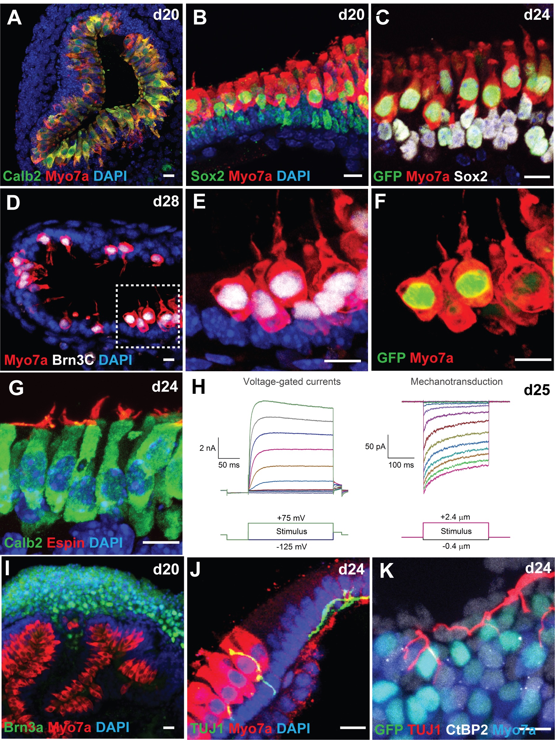
CHIR-treated aggregates give rise to inner ear organoids harboring mechanosensitive hair cells
(A-B) Co-localization of two hair cell markers Calb2 and Myo7a (A) or Sox2 and Myo7a (B) in cells lining the luminal surface of a vesicle.
(C) Atoh1/nGFP+ cells also express Myo7a and Sox2.
(D-F) Cells expressing Brn3C, Myo7a, and Atoh1/nGFP exhibit flask-like morphology with a hair bundle on their apical surface characteristic of vestibular hair cells. Some Myo7a+ cells in day 28 samples have faint Atoh1/nGFP expression (arrows), suggesting that these cells are more mature hair cells than cells expressing strong Atoh1/nGFP expression.
(G) The hair bundle marker Espin was observed on the apical surface of Calb2+ hair cells.
(H) Representative voltage-gated currents and mechanosensitive currents recorded from day 25 Atoh1/nGFP+ cells in response to voltage injections and hair bundle deflections, respectively.
(I) A cluster of Brn3A+ neuronal cell bodies were located near Myo7a+ hair cells.
(J) A TUJ1+ neural processes extend and contact Myo7a+ hair cells. (K) The ribbon synapse marker CtBP2 was associated with Myo7a+ cells and TUJ1+ processes.
Scale bars, 10 μm (A-G, I-K).
Reference
DeJonge RE, Liu XP, Deig CR, Heller S, Koehler KR & Hashino E. (2016). Modulation of Wnt Signaling Enhances Inner Ear Organoid Development in 3D Culture. PLoS ONE , 11, e0162508. PMID: 27607106 DOI.
Copyright
© 2016 DeJonge et al. This is an open access article distributed under the terms of the Creative Commons Attribution License, which permits unrestricted use, distribution, and reproduction in any medium, provided the original author and source are credited.
Fig 5. Journal.pone.0162508.g005.jpg
https://doi.org/10.1371/journal.pone.0162508.g005
Cite this page: Hill, M.A. (2024, April 25) Embryology Organoids - inner ear 01.jpg. Retrieved from https://embryology.med.unsw.edu.au/embryology/index.php/File:Organoids_-_inner_ear_01.jpg
- © Dr Mark Hill 2024, UNSW Embryology ISBN: 978 0 7334 2609 4 - UNSW CRICOS Provider Code No. 00098G
File history
Click on a date/time to view the file as it appeared at that time.
| Date/Time | Thumbnail | Dimensions | User | Comment | |
|---|---|---|---|---|---|
| current | 06:16, 30 July 2019 | | 1,796 × 2,396 (1.07 MB) | Z8600021 (talk | contribs) | ==CHIR-treated aggregates give rise to inner ear organoids harboring mechanosensitive hair cells== (A-B) Co-localization of two hair cell markers Calb2 and Myo7a (A) or Sox2 and Myo7a (B) in cells lining the luminal surface of a vesicle. (C) Atoh1/nG... |
You cannot overwrite this file.
File usage
The following page uses this file: