File:Odgers1941 text-fig02.jpg

Original file (1,000 × 371 pixels, file size: 24 KB, MIME type: image/jpeg)
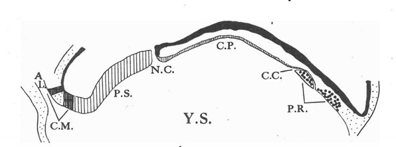
Text-fig. 2. Longitudinal section through the embryonal axis
A vertical longitudinal section through the embryonal axis (x 75) drawn diagrammatically to scale and idealized to permit a representation of all the axial structures in the same section. AL. allantois. C.M. cloacal membrane. P.S. primitive streak. N.C.neurenteric canal. C.P. chorda plate. P.R. prochordal plate. C.G.remains of chorda canal. Y.S. yolk sac.
Reference
Odgers PN. A presomite human embryo with a neurenteric canal (embryo R.S.). (1941) J. Anat., 75(4): 381-388.3. PMID 17104868
Cite this page: Hill, M.A. (2024, April 24) Embryology Odgers1941 text-fig02.jpg. Retrieved from https://embryology.med.unsw.edu.au/embryology/index.php/File:Odgers1941_text-fig02.jpg
- © Dr Mark Hill 2024, UNSW Embryology ISBN: 978 0 7334 2609 4 - UNSW CRICOS Provider Code No. 00098G
File history
Click on a date/time to view the file as it appeared at that time.
| Date/Time | Thumbnail | Dimensions | User | Comment | |
|---|---|---|---|---|---|
| current | 16:52, 22 October 2017 | 1,000 × 371 (24 KB) | Z8600021 (talk | contribs) | ===Reference=== {{Ref-Odgers1941}} {{Footer}} |
You cannot overwrite this file.
File usage
The following page uses this file: