File:Odgers1941 plate01.jpg

Original file (1,280 × 1,677 pixels, file size: 200 KB, MIME type: image/jpeg)
Plate 1
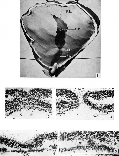
Fig. 1. Photograph of the dorsal aspect of a reconstruction model of the embryonal disc (x 75). The amniotic sac has been removed and the allantois dissected out of the body stalk, while the differentportionsoftheaxialformationsareportrayedonthesurface. P.S.primitivestreak. N.C.neurentericcanal. C.P. chorda plate. P.R.prochordal plate.
Fig. 2. A section (x 250) through the primitive streak. It shows the activity of the entoderm whichatX may be contributing to the mesoderm.
Fig. 3. A section (x250) through the neurenteric canal, N.C. C.P. indicates the commencement of the chordplate. Y.S. yolk sac.
Fig. 4. A typical section (x250) of the chordaplate,C.P.
Fig. 5. A section (x370) showing a mass of cels at X contiguous ventrally with the dorsal surface of the chord plate and dorsally with the ectoderm.
Reference
Odgers PN. A presomite human embryo with a neurenteric canal (embryo R.S.). (1941) J. Anat., 75(4): 381-388.3. PMID 17104868
Cite this page: Hill, M.A. (2024, April 25) Embryology Odgers1941 plate01.jpg. Retrieved from https://embryology.med.unsw.edu.au/embryology/index.php/File:Odgers1941_plate01.jpg
- © Dr Mark Hill 2024, UNSW Embryology ISBN: 978 0 7334 2609 4 - UNSW CRICOS Provider Code No. 00098G
File history
Click on a date/time to view the file as it appeared at that time.
| Date/Time | Thumbnail | Dimensions | User | Comment | |
|---|---|---|---|---|---|
| current | 16:28, 22 October 2017 |  | 1,280 × 1,677 (200 KB) | Z8600021 (talk | contribs) | |
| 16:27, 22 October 2017 |  | 1,665 × 2,345 (462 KB) | Z8600021 (talk | contribs) |
You cannot overwrite this file.
File usage
The following page uses this file: