File:Odgers1938 plate02.jpg
Original file (1,673 × 2,500 pixels, file size: 986 KB, MIME type: image/jpeg)
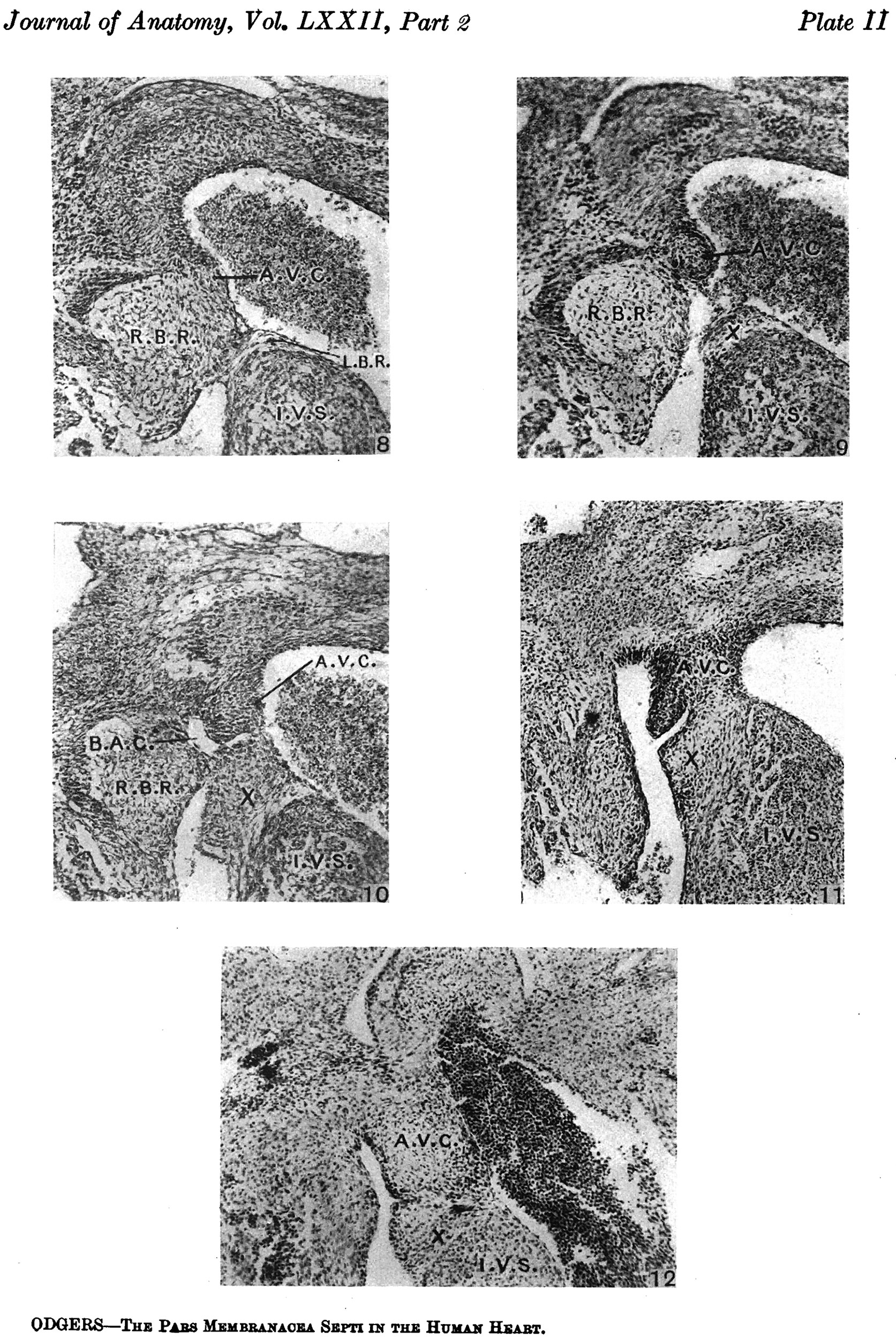
Plate II
Abbreviations
- R.B.R. right bulbar ridge.
- L.B.R. left bulbar ridge.
- R.S.T. right superior tubercle of the A.-V. cushions.
- R.I.T. right inferior tubercle of the A.-V. cushions.
- I.V.S. interventricular septum.
- A.V.C. auriculoventricular cushions.
- B.A.C. bulbo-auricular channel.
Figs. 8-10. These are microphotographs of sections from a 155 mm. embryo (x 80). Fig. 8 is immediately cranial to the interventricular foramen and shows that in the proximal bulbar septum the right, R.B.R., and left, L.B.R., bulbar ridges can still be distinguished. The right one is fused with the right superior tubercle of the A.-V. cushions, A. 17.0., while the lefi one joins the interventricular septum, I . V.S. Fig. 9 is a section through the centre of the foramen and shows the septum, I. 17.8., capped with cushion tissue, X. The latter, as is seen in Fig. 10, which is the fourth section caudal to the level of the foraminal closure, joins the A.-V. cushion. A. V.C’., on its left side but is still separated from it on the right by a notch, which appears as a diverticulum from the anterior end of the inferior portion of the bulbo-auricular channel, B.A.C'.
Fig. 11. This is a section through the heart of a 17 mm. embryo ( x 80) five sections below the closing foramen. It shows the A.-V. cushion, A.V.0., separated, as in the last figure, by a notch on the right side from the cushion tissue, X, which has proliferated from it to join the interventricular septum, I. 17.18’.
Fig. 12. This is a section from a 17-5 mm. embryo ( x 80) and shows a notch in the same situation as that seen above, but now it is much less well marked.
Fig 8.
Fig 9.
Fig 10.

Fig 11.

Fig 12.


| Historic Disclaimer - information about historic embryology pages |
|---|
 |
Reference
Odgers PN. The development of the pars membranacea septi in the human heart. (1938) J Anat. 72(2): 247-59. https://www.ncbi.nlm.nih.gov/pubmed/17104688 PMID 17104688]
Cite this page: Hill, M.A. (2024, April 19) Embryology Odgers1938 plate02.jpg. Retrieved from https://embryology.med.unsw.edu.au/embryology/index.php/File:Odgers1938_plate02.jpg
- © Dr Mark Hill 2024, UNSW Embryology ISBN: 978 0 7334 2609 4 - UNSW CRICOS Provider Code No. 00098G
File history
Click on a date/time to view the file as it appeared at that time.
| Date/Time | Thumbnail | Dimensions | User | Comment | |
|---|---|---|---|---|---|
| current | 10:32, 19 January 2016 | | 1,673 × 2,500 (986 KB) | Z8600021 (talk | contribs) |
You cannot overwrite this file.
File usage
The following page uses this file: