File:Odgers1938 plate01.jpg

Original file (1,854 × 2,500 pixels, file size: 1.09 MB, MIME type: image/jpeg)
Plate I
Abbreviations
- R.B.R. right bulbar ridge.
- L.B.R. left bulbar ridge.
- R.S.T. right superior tubercle of the A.-V. cushions.
- R.I.T. right inferior tubercle of the A.-V. cushions.
- I.V.S. interventricular septum.
- A.V.C. auriculoventricular cushions.
- B.A.C. bulbo-auricular channel.
Fig. 1. A section immediately caudal to the interventricular foramen in an 11-2 mm. embryo ( x 80) to show that a tongue-shaped proliferation from the A.-V. cushion, R.I.T., is making contact with the right side of the interventricular septum, I. V.S., ventral to its dorsal border.
Fig. 2. A section through the interventricular foramen in an 11-4 mm. embryo ( x 80). It shows the right bulbar ridge, R.B.R., in contact with the right superior tubercle of the A.—V. cushions, R.S.T., to form the bulbo-auricular channel, B.A.0. R.I.TL marks the inferior A.-V. tubercle proliferating cranialwards.
Figs. 3-5. Sections through this region of the heart in a 12-5 mm. embryo ( x 80). Fig. 3 shows the interventricular septum, I. V.S., capped by cushion tissue (X), which is in contact with the right bulbar ridge, R.B.R. In Fig. 4, which is the next section below Fig. 3, this tissue X is connected with both the right bulbar ridge, R.B.R., and with the A.-V. cushions, A.V.0. Fig. 5 is a section three below the last and shows the proliferating tissue joining the A.-V. cushions, A.V.0. The junction between the two is marked on either side by a notch. The bulbo-auricular channel, B.A.C'., appears in Fig. 3 as a. narrow cleft between the right bulbar ridge, R.B.R., and the A.-V. cushions, A. V.0.: in Fig. 4 this channel is better marked, while in Fig. 5 it is seen to be continuous with the right auricle.
Figs. 6-7. These are sections through the heart of a 14-5 mm. embryo ( x 80). In Fig. 6, which is through the centre of the interventricular foramen, the A.-V. cushion, A. V.0'., is seen to be proliferating, while similar cushion tissue, X, caps the interventricular septum, I. 17.8. In Fig. 7 the proliferating cushion tissue is making contact with that on the septum, X, while its right border is joined by the right bulbar ridge, R.B.R., as it encloses the bulbo-auricular channel, BA .0.
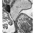
Fig 1.
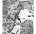
Fig 2.

Fig 3.
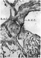
Fig 4.
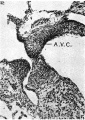
Fig 5.

Fig 6.
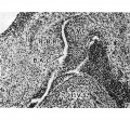
Fig 7.


| Historic Disclaimer - information about historic embryology pages |
|---|
 |
Reference
Odgers PN. The development of the pars membranacea septi in the human heart. (1938) J Anat. 72(2): 247-59. https://www.ncbi.nlm.nih.gov/pubmed/17104688 PMID 17104688]
Cite this page: Hill, M.A. (2024, April 25) Embryology Odgers1938 plate01.jpg. Retrieved from https://embryology.med.unsw.edu.au/embryology/index.php/File:Odgers1938_plate01.jpg
- © Dr Mark Hill 2024, UNSW Embryology ISBN: 978 0 7334 2609 4 - UNSW CRICOS Provider Code No. 00098G
File history
Click on a date/time to view the file as it appeared at that time.
| Date/Time | Thumbnail | Dimensions | User | Comment | |
|---|---|---|---|---|---|
| current | 10:27, 19 January 2016 | | 1,854 × 2,500 (1.09 MB) | Z8600021 (talk | contribs) |
You cannot overwrite this file.
File usage
The following page uses this file: