File:O'Rahilly1956 plate01.jpg
Original file (1,599 × 2,032 pixels, file size: 293 KB, MIME type: image/jpeg)
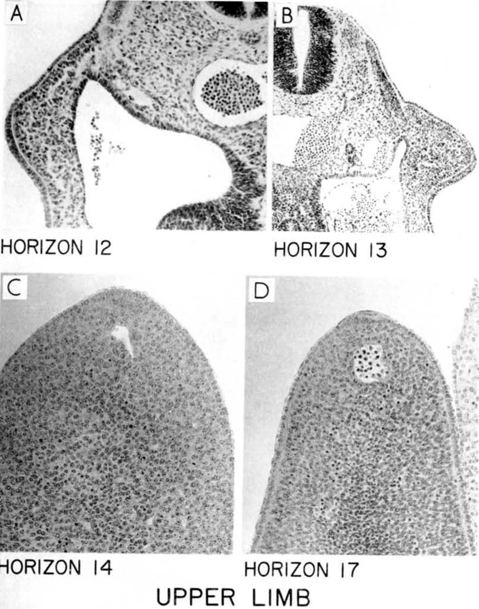
Plate 1. Upper Limb
Magnifications given in Plates 1 and 2 are the original ones used in taking the photomicrographs.
FIG. A. Horizon 12. Embryo No. 5923. 4 mm. 2-3-2. x 200. The upper limb bud projects at the left-hand side of the photomicrograph. The ectodermal thickening can be seen on the ventral surface and lateral margin of the limb bud. The extent of the thickening in this embryo can be assessed from Text-fig. 1.
FIG. B. Horizon 13. Embryo No. 8066. 53 mm. 6-4-4. x 100. The upper limb bud projects at the right-hand side of the photomicrograph. The ectodermal thickening shows several layers of nuclei.
FIG. C. Horizon 14. Embryo No. 8552. 65 mm. 8-3-4. x200. The lateral portion of the ecto- dermal thickening forms a prominent elevation, the ectodermal ridge, which occupies the ventro- lateral or pre-axial border of the limb. The marginal vein can be seen adjacent to the ridge.
FIG. D. Horizon 17. Embryo No. 8998. 11 mm. 21-1-3. x200. The ectodermal ridge forms the margin of the digital plate.
Cite this page: Hill, M.A. (2024, April 24) Embryology O'Rahilly1956 plate01.jpg. Retrieved from https://embryology.med.unsw.edu.au/embryology/index.php/File:O%27Rahilly1956_plate01.jpg
- © Dr Mark Hill 2024, UNSW Embryology ISBN: 978 0 7334 2609 4 - UNSW CRICOS Provider Code No. 00098G
File history
Click on a date/time to view the file as it appeared at that time.
| Date/Time | Thumbnail | Dimensions | User | Comment | |
|---|---|---|---|---|---|
| current | 14:57, 2 July 2018 | | 1,599 × 2,032 (293 KB) | Z8600021 (talk | contribs) | |
| 14:56, 2 July 2018 |  | 1,713 × 2,290 (281 KB) | Z8600021 (talk | contribs) |
You cannot overwrite this file.
File usage
There are no pages that use this file.