File:Noback1943-plate02.jpg

Original file (1,000 × 1,436 pixels, file size: 198 KB, MIME type: image/jpeg)
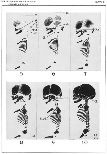
Plate 2
Side View drawings of six stages in the development of the human prenatal and cireumnntnl ossoous skeleton. C71-own-riunp lengths are held constant in all figures.
Note: (1) the maturation of the facial bones, ribs nnd clavicle in the stage depicted in figure 3, (2) maturation of the calvarial bones in the stage depicted by figure 5, (3) the central plate, the open reticular zone and the marginal zone of the calvarial bones, and (4) the relatively slow growth of most of the cartilage bones as exemplified by the supruoeciptal bone and the 1'eln.tive1y rapid growth of the membrane bones as exemplified by the interparietal bone.
| CR LENGTH | CH LENGTH | APPROXIMATE AGE | |
|---|---|---|---|
| Figure 5 | 38 mm. | 64 days | |
| Figure 6 | 58 mm. | 75 mm. | 78 days |
| Figure 7 | 88 mm. | 125 mm. | 3.25 lunar months |
| Figure 8 | 105 mm. | 156 mm. | 3.5 lunar months |
| Figure 9 | 139 mm. | 205 mm. | 4.25 lunar months |
| Figure 10 | 330 mm. | 479 mm. | 10 lunar months |
Legend: A., alisphenoid bone; B., basioeoipital bone; Bs., hasisphenoid bone; 0., crown; E., exoccipital bone; I., interparietal bone; Is., ischinm; P., petrosum; Pu., pubis; S., supraoceipital bone; S.M., sternal center of the manubrium; S.P., site of the pertosum; T.A., tympanic annulus.
| Historic Disclaimer - information about historic embryology pages |
|---|
| Pages where the terms "Historic" (textbooks, papers, people, recommendations) appear on this site, and sections within pages where this disclaimer appears, indicate that the content and scientific understanding are specific to the time of publication. This means that while some scientific descriptions are still accurate, the terminology and interpretation of the developmental mechanisms reflect the understanding at the time of original publication and those of the preceding periods, these terms, interpretations and recommendations may not reflect our current scientific understanding. (More? Embryology History | Historic Embryology Papers) |
Reference
Noback CR. Some gross structural and quantitative aspects of the developmental anatomy of the human embryonic, fetal and circumnatal skeleton. (1943) The Anat. Rec. 87: 29–51.
Cite this page: Hill, M.A. (2024, April 19) Embryology Noback1943-plate02.jpg. Retrieved from https://embryology.med.unsw.edu.au/embryology/index.php/File:Noback1943-plate02.jpg
- © Dr Mark Hill 2024, UNSW Embryology ISBN: 978 0 7334 2609 4 - UNSW CRICOS Provider Code No. 00098G
File history
Click on a date/time to view the file as it appeared at that time.
| Date/Time | Thumbnail | Dimensions | User | Comment | |
|---|---|---|---|---|---|
| current | 08:53, 15 November 2015 |  | 1,000 × 1,436 (198 KB) | Z8600021 (talk | contribs) |
You cannot overwrite this file.
File usage
The following page uses this file:
