File:Neutrophil chasing bacteria.jpg
Neutrophil_chasing_bacteria.jpg (640 × 480 pixels, file size: 56 KB, MIME type: image/jpeg)
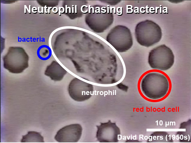
Neutrophil Chasing Bacteria
Scale bar = 10 microns |
|
This still image was taken from a 16-mm movie made in the 1950s by the late David Rogers at Vanderbilt University. Labels have been aded and the image is used as the link icon for the associated movie. Click on Page or Play above to see the original movie.
About the Movie
This video is taken from a 16-mm movie made in the 1950s by the late David Rogers at Vanderbilt University. It was given to Thomas P. Stossel via Dr. Victor Najjar, Professor Emeritus at Tufts University Medical School and a former colleague of Rogers.
It depicts a human polymorphonuclear leukocyte (neutrophil) on a blood film, crawling among red blood cells, notable for their dark color and principally spherical shape. The neutrophil is "chasing" Staphylococcus aureus microorganisms, added to the film.
The chemoattractant derived from the microbe is unclear but may be complement fragment C5a, generated by the interaction of antibodies in the blood serum with the complement cascade, and/or bacterial N-formyl peptides. Blood platelets adherent to the underlying glass are also visible. Notable is the characteristic asymmetric shape of the crawling neutrophil with an organelle-excluding leading lamella and a narrowing at the opposite end culminating in a "tail" that the cell appears to drag along. Contraction waves are visible along the surface of the moving cell as it moves forward in a gliding fashion. As the neutrophil relentlessly pursues the microbe it ignores the red cells and platelets. However, its leading edge is sufficiently stiff (elastic) to deform and displace the red cells it bumps into.
The internal contents of the neutrophil also move, and granule motion is particularly dynamic near the leading edge. These granules only approach the cell surface membrane when the cell changes direction and redistributes its peripheral "gel." After the neutrophil has engulfed the bacterium, note that the cell's movements become somewhat more jerky, and that it begins to extend more spherical surface projections. These bleb-like protruberances resemble the blebs that form constitutively in the M2 melanoma cells missing the actin filament crosslinking protein filamin-1 (ABP-280) and may be telling us something about the mechanism of membrane protrusion.
Text: Thomas P. Stossel (Brigham and Women's Hospital and Harvard Medical School), June 22, 1999
File history
Click on a date/time to view the file as it appeared at that time.
| Date/Time | Thumbnail | Dimensions | User | Comment | |
|---|---|---|---|---|---|
| current | 16:30, 1 February 2015 | | 640 × 480 (56 KB) | Z8600021 (talk | contribs) | |
| 11:25, 17 January 2012 |  | 396 × 296 (16 KB) | S8600021 (talk | contribs) | ==Neutrophil Chasing Bacteria== {| | 300px | * <font color=blue>'''blue ring - bacteria'''</font> * '''white ring - white blood cell''' * <font color=red>'''red ring - red blood cell'''</font> Scale bar = 10 microns |} This still i |
You cannot overwrite this file.
File usage
The following 5 pages use this file: