File:Nelsen1953 fig004.jpg

Original file (1,200 × 960 pixels, file size: 200 KB, MIME type: image/jpeg)
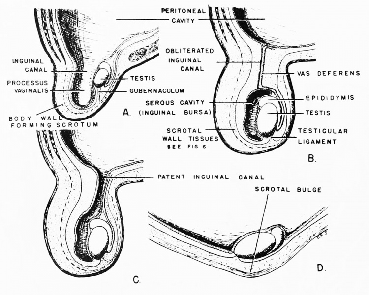
Fig. 4. Diagrammatic drawings portraying the relationship of the testis to the processus vaginalis (peritoneal evagination) and the scrotum
The testis is at all times retroperitoneal, i.e., outside the peritoneal cavity and membrane.
(A) Earlier stage of testicular descent at the time the testis is moving downward into the scrotum.
(B) Position of the testis at the end of its scrotal journey in a form possessing permanent descent of the testis, e.g., man, dog, etc.
(C) Testis-peritoneal relationship in a form which does not have a permanent descent of the testis - the testis is withdrawn into the peritoneal cavity at the termination of each breeding season. Shortly before the onset of the breeding period or "rut," the testis once again descends into the scrotum, e.g., ground hog.
(D) Position of testis in relation to body wall and peritoneum in the mole, shrew, and hedgehog in which there is no true scrotum. The testis bulges outward, pushing the body wall before it during the breeding season. As the testis shrinks following the season of rut, the bulge in the body wall recedes. True also of bat, Myotis.
Reference
Nelsen OE. Comparative embryology of the vertebrates (1953) Mcgraw-Hill Book Company, New York.
Cite this page: Hill, M.A. (2024, April 19) Embryology Nelsen1953 fig004.jpg. Retrieved from https://embryology.med.unsw.edu.au/embryology/index.php/File:Nelsen1953_fig004.jpg
- © Dr Mark Hill 2024, UNSW Embryology ISBN: 978 0 7334 2609 4 - UNSW CRICOS Provider Code No. 00098G
File history
Click on a date/time to view the file as it appeared at that time.
| Date/Time | Thumbnail | Dimensions | User | Comment | |
|---|---|---|---|---|---|
| current | 14:25, 26 October 2016 |  | 1,200 × 960 (200 KB) | Z8600021 (talk | contribs) | |
| 14:25, 26 October 2016 |  | 1,878 × 1,915 (568 KB) | Z8600021 (talk | contribs) |
You cannot overwrite this file.
File usage
The following 2 pages use this file: