File:Nanagas1925-fig01c.jpg
Original file (600 × 765 pixels, file size: 34 KB, MIME type: image/jpeg)
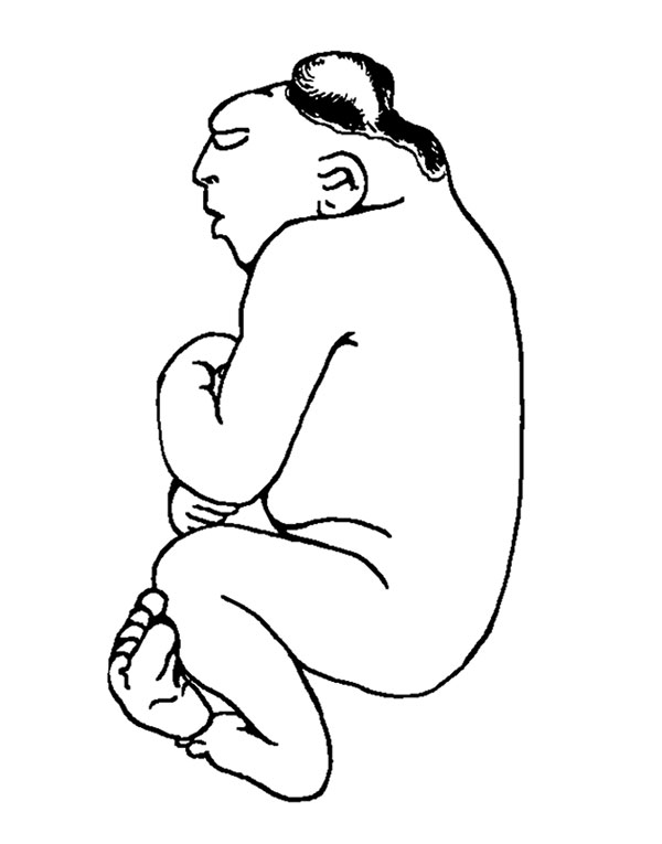
Fig. 1c. Microcephalic Acrauius
This is a condition of acrania with the persistence of a diminutive and imperfectly developed brain. The brain tissue is represented by a soft, flabby mass of variable size which occupies the flattened base of the vaultless cranium. A meningeal-like membrane covers this structure in all cases. In some instances, fluid is present within this tumor-like mass, giving it the form of a cyst with walls of varying thickness. Such cases would more properly come within the subclassification of hydromicrocephalus. There were seven cases of microcephalic acrania in the series (12.3 per cent).
| Historic Disclaimer - information about historic embryology pages |
|---|
 |
- Links: Fig 1. Anencephalus types | Fig 1a. anencephalic acranius | Fig 1b. anencephalic craniorhachischisis | Fig 1c. microcephalic acrauius | Fig 1d. microcephalic craniorhachischisis | Fig 1e. exocephalic acranius | Fig 16. anencephalic and normal fetuses | Historic Embryology Papers | Neural Abnormalities | Folic Acid and Neural Tube Defects | Skull Development
Reference
Nañagas JC. A comparison of the growth of the body dimensions of anencephalic human fetuses with normal fetal growth as determined by graphic analysis and empirical formulae. (1925) American J. Anatomy. 455-494.
Cite this page: Hill, M.A. (2024, April 18) Embryology Nanagas1925-fig01c.jpg. Retrieved from https://embryology.med.unsw.edu.au/embryology/index.php/File:Nanagas1925-fig01c.jpg
- © Dr Mark Hill 2024, UNSW Embryology ISBN: 978 0 7334 2609 4 - UNSW CRICOS Provider Code No. 00098G
File history
Click on a date/time to view the file as it appeared at that time.
| Date/Time | Thumbnail | Dimensions | User | Comment | |
|---|---|---|---|---|---|
| current | 12:22, 16 September 2015 | | 600 × 765 (34 KB) | Z8600021 (talk | contribs) | {{Nanagas1925 figures}} |
You cannot overwrite this file.