File:Muscle- centrosome protein localizes cytoplasmic site nuclear envelope.jpg

Original file (1,000 × 1,840 pixels, file size: 293 KB, MIME type: image/jpeg)
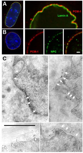
The centrosome protein PCM-1 localizes to dense structures on the cytoplasmic site of the nuclear envelope.
(A) Deconvolved image of a nucleus from a differentiated H-2Kb-tsA58 cell, expressing GFP-lamin A (green), and stained for PCM-1 (red) and DNA (blue).
(B) Nucleus from a differentiated H-2Kb-tsA58 cell, stained for PCM-1 (red), and for nuclear pore complex proteins (NPC, green). Selected areas of the nuclei in (A) and (B) are shown enlarged on the right.
(C) Immuno-electron microscopy of cryosections of differentiated C2C12 cells. Different views of cross-sections of the nucleus are shown. PCM-1 is labelled with antibody and protein A, coupled to 10 nm gold. White arrows indicate the outline of a layer of electron-dense material at the outer nuclear surface.
Bars in (B) and (C), 1 μm.
http://www.biomedcentral.com/1471-2121/10/28/figure/F4
Original Figure cropped and resolution changed.
Centrosome proteins form an insoluble perinuclear matrix during muscle cell differentiation. Srsen V, Fant X, Heald R, Rabouille C, Merdes A. BMC Cell Biol. 2009 Apr 21;10:28. PMID: 19383121 | BMC Cell Biol.
© 2009 Srsen et al; licensee BioMed Central Ltd.
This is an Open Access article distributed under the terms of the Creative Commons Attribution License (http://creativecommons.org/licenses/by/2.0), which permits unrestricted use, distribution, and reproduction in any medium, provided the original work is properly cited.
File history
Click on a date/time to view the file as it appeared at that time.
| Date/Time | Thumbnail | Dimensions | User | Comment | |
|---|---|---|---|---|---|
| current | 12:31, 1 May 2010 |  | 1,000 × 1,840 (293 KB) | S8600021 (talk | contribs) | The centrosome protein PCM-1 localizes to dense structures on the cytoplasmic site of the nuclear envelope. (A) Deconvolved image of a nucleus from a differentiated H-2Kb-tsA58 cell, expressing GFP-lamin A (green), and stained for PCM-1 (red) and DNA (b |
You cannot overwrite this file.
File usage
There are no pages that use this file.