File:Mouse zygote pronuclei 03.jpg

Original file (1,200 × 586 pixels, file size: 66 KB, MIME type: image/jpeg)
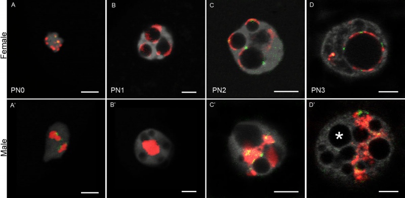
Mouse Zygote Male and Female Pronulei
3D-FISH images obtained on early 1-cell stage embryos with pericentromeric and centromeric probes. Different reorganization within the male and female pronuclei from PN0 to PN5.
Pericentromeres after fertilization
- female pronucleus (fPN; maternally inherited genome) - organized rapidly around the NPBs
- male pronucleus (mPN; paternally inherited genome) - remained associated together less unorganized masses located in the centre
- Links: Zygote | Mouse Development
Reference
<pubmed>23095683</pubmed>| BMC Dev Biol.
Copyright
© 2012 Aguirre Lavin et al.; licensee BioMed Central Ltd. This is an Open Access article distributed under the terms of the Creative Commons Attribution License ( http://creativecommons.org/licenses/by/2.0), which permits unrestricted use, distribution, and reproduction in any medium, provided the original work is properly cited.
Additional Figure 1. 1471-213x-12-30-s1 resized. Text modified from paper and legend.
File history
Click on a date/time to view the file as it appeared at that time.
| Date/Time | Thumbnail | Dimensions | User | Comment | |
|---|---|---|---|---|---|
| current | 11:58, 29 December 2012 |  | 1,200 × 586 (66 KB) | Z8600021 (talk | contribs) | 3D-FISH images obtained on early 1-cell stage embryos with pericentromeric and centromeric probes. :'''Links:''' Zygote | Mouse Development ===Reference=== <pubmed>23095683</pubmed>| [http://www.biomedcentral.com/1471-213X/12/30 BMC Dev Biol.] |
You cannot overwrite this file.
File usage
There are no pages that use this file.