File:Mouse seminiferous tubule EM.jpg

Original file (1,152 × 862 pixels, file size: 352 KB, MIME type: image/jpeg)
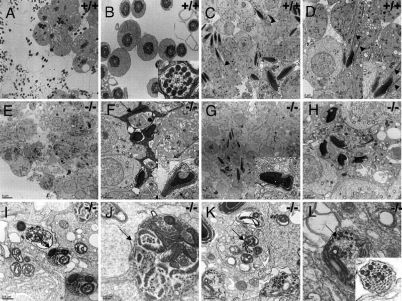
Testicular ultrastructure in adult wild-type and Meig1 mutant mice.
Representative transmission electronic microscopy images from an adult wild-type mouse (A–D) and a Meig1 homozygous mutant mouse (E–L).
A and B show normal spermatogenesis and axoneme structure in wild-type testes, C and D shows normal condensing spermatids heads and manchette structure (arrow heads) in wild-type testes.
E shows failure of spermatogenesis as evaluated by absence of sperm in the lumen of seminiferous tubule. F represents highly condensed Sertoli cell cytoplasm (arrow). G and H represent some condensing spermatids that lack manchette structure as seen in the wild-type testes (C and D). Inserts in F and G represent two deformed sperm heads.
I–L and inserts show disorganized flagella. Note that the flagella components, such as microtubules and outer dense fibers seem to be made normally, but are not assembled correctly into flagella. Some flagella contain multiple axonemal structure (J). Arrows point to disorganized flagella.
http://www.pnas.org/content/106/40/17055/F3.expansion.html
http://www.ncbi.nlm.nih.gov/pubmed/19805151
http://www.pnas.org/content/106/40/17055.abstract
MEIG1 is essential for spermiogenesis in mice, Zhibing Zhang, Xuening Shen, David R. Gude, Bonney M. Wilkinson, Monica J. Justice, Charles J. Flickinger, John C. Herr, Edward M. Eddy, and Jerome F. Strauss, III
- "Spermatogenesis can be divided into three stages: spermatogonial mitosis, meiosis of spermatocytes, and spermiogenesis. During spermiogenesis, spermatids undergo dramatic morphological changes including formation of a flagellum and chromosomal packaging and condensation of the nucleus into the sperm head. The genes regulating the latter processes are largely unknown. ...Our findings reveal a critical role for the MEIG1/PARCG partnership in manchette structure and function and the control of spermiogenesis."
Anyone may, without requesting permission, use original figures or tables published in PNAS for noncommercial and educational use (i.e., in a review article, in a book that is not for sale) provided that the original source and the applicable copyright notice are cited.
File history
Click on a date/time to view the file as it appeared at that time.
| Date/Time | Thumbnail | Dimensions | User | Comment | |
|---|---|---|---|---|---|
| current | 09:44, 7 October 2009 |  | 1,152 × 862 (352 KB) | S8600021 (talk | contribs) | Testicular ultrastructure in adult wild-type and Meig1 mutant mice. Representative transmission electronic microscopy images from an adult wild-type mouse (A–D) and a Meig1 homozygous mutant mouse (E–L). A and B show normal spermatogenesis and axo |
You cannot overwrite this file.
File usage
There are no pages that use this file.