File:Mouse primitive node cilia.jpg

Original file (592 × 981 pixels, file size: 130 KB, MIME type: image/jpeg)
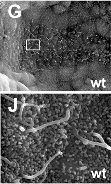
Mouse primitive node cilia (E7.5–8.0)
Extract from Figure 6: Nodal cilia are present but abnormal in Tbx6 mutant embryos. (A–F) Confocal images through the node of wild type (wt) and Tbx6 mutant −/−) embryos stained with anti-acetylated tubulin with a Hoechst nuclear counterstain.
Cilia visible on the ventral surface are filamentous in wild type but are short with bulbous tips in the mutant. (G–L) SEM images of the node and nodal cilia in wt and −/− nodes (G,J) Normal, long filamentous cilia in wild type nodes. (H,K) Mutant node showing cilia with blebs and bulbous tips typical of most mutants. (I,L) Atypical mutant node with short cilia and an abnormally shaped node with large visceral endoderm-like cells within the node (arrow). The boxes in G–I correspond to the higher magnification views in J–L.
http://www.plosone.org/article/info:doi%2F10.1371%2Fjournal.pone.0002511
Citation: Hadjantonakis A-K, Pisano E, Papaioannou VE (2008) Tbx6 Regulates Left/Right Patterning in Mouse Embryos through Effects on Nodal Cilia and Perinodal Signaling. PLoS ONE 3(6): e2511. doi:10.1371/journal.pone.0002511
Editor: Thomas Zwaka, Baylor College of Medicine, United States of America
Received: March 3, 2008; Accepted: May 24, 2008; Published: June 25, 2008
Copyright: © 2008 Hadjantonakis et al. This is an open-access article distributed under the terms of the Creative Commons Attribution License, which permits unrestricted use, distribution, and reproduction in any medium, provided the original author and source are credited.
File history
Click on a date/time to view the file as it appeared at that time.
| Date/Time | Thumbnail | Dimensions | User | Comment | |
|---|---|---|---|---|---|
| current | 09:39, 15 April 2010 | | 592 × 981 (130 KB) | S8600021 (talk | contribs) | Mouse primitive node cilia (E7.5–8.0) Extract from Figure 6: Nodal cilia are present but abnormal in Tbx6 mutant embryos. (A–F) Confocal images through the node of wild type (wt) and Tbx6 mutant −/−) embryos stained with anti-acetylated tubulin w |
You cannot overwrite this file.
File usage
There are no pages that use this file.