File:Mouse inner ear.jpg
Mouse_inner_ear.jpg (478 × 508 pixels, file size: 47 KB, MIME type: image/jpeg)
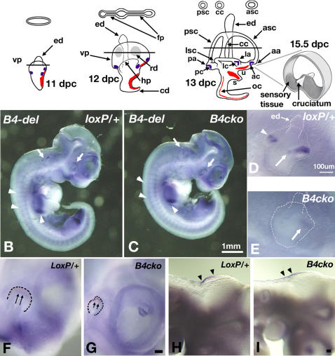
Schematic representations of mouse inner ear development from 11.5 to 13 dpc
(A) Upper panel shows schematic cross-sections through the prospective or definitive anterior and posterior canals at the level of the lines. Blue marks the three Bmp4-positive presumptive cristae, while red marks the other three sensory tissues-the maculae utriculi and sacculi, and the organ of Corti. An enlargement of a mature anterior crista at 15.5 dpc or later is shown. (B–I) Inner ear phenotypes of Bmp4 conditional null embryos. Wholemount in situ hybridization of Bmp4loxP/+ (B,D,F,H) and Foxg1cre/+; Bmp4loxP/Tm1 (B4cko, C,E,G,I) embryos at 10.5 dpc hybridized with Bmp4 RNA probe specific for exons 3 and 4 (B4-del).
(B, C) Arrows point to the down-regulation of Bmp4 expression in the eyes and otocysts of Foxg1cre/+; Bmp4loxP/Tm1 (C), compared to Bmp4loxP/+ embryos (B). Arrowheads point to unaffected Bmp4 expression in limb buds and somites.
(D) and (E) are higher magnifications of the otocysts shown in (B) and (C), respectively. Arrow and arrowhead in (D) point to Bmp4 hybridization signals in the anterior streak (encompassing anterior and lateral cristae) and the posterior crista of the otocyst, respectively. An arrow in (E) points to the residual Bmp4 expression in the anterior streak of Foxg1cre/+; Bmp4loxP/Tm1 embryos. (F–I) Higher magnifications of Bmp4 expression domains in the eyes (F, G) and hindbrain (H,I) in Bmp4loxP/+ (F, H) and Foxg1cre/+; Bmp4loxP/Tm1 (G, I) embryos. Arrows point to the reduction of Bmp4 expression, and the malformation of the eyes, whereas arrowheads point to the normal Bmp4 expression in the hindbrain. Scale bar in (C) applies to (B); scale bars in (D), (G) and (I) equal 100μm and apply to (E), (F), and (H), respectively.
Abbrevations: aa, anterior ampulla; ac, anterior crista; asc, anterior semicircular canal; cc, common crus; cd, cochlear duct; ed, endolymphatic duct; fp, fusion plate; hp, horizontal canal pouch; la, lateral ampulla; lc, lateral crista; lsc, lateral semicircular canal; oc, organ of Corti; pa, posterior ampulla; pc, posterior crista; psc, posterior semicircular canal; rd, resorption domain; s, saccule; u, utricle; vp, vertical canal pouch.
Reference
PLoS Genet. 2008 April; 4(4): e1000050. Published online 2008 April 11. doi: 10.1371/journal.pgen.1000050. http://www.pubmedcentral.nih.gov/articlerender.fcgi?artid=2274953
Copyright This is an open-access article distributed under the terms of the Creative Commons Public Domain declaration which stipulates that, once placed in the public domain, this work may be freely reproduced, distributed, transmitted, modified, built upon, or otherwise used by anyone for any lawful purpose.
Original Image Name: Pgen.1000050.g001.jpg
File history
Click on a date/time to view the file as it appeared at that time.
| Date/Time | Thumbnail | Dimensions | User | Comment | |
|---|---|---|---|---|---|
| current | 21:50, 24 September 2009 |  | 478 × 508 (47 KB) | S8600021 (talk | contribs) | Schematic representations of mouse inner ear development from 11.5 to 13 dpc. (A) Upper panel shows schematic cross-sections through the prospective or definitive anterior and posterior canals at the level of the lines. Blue marks the three Bmp4-positive |
You cannot overwrite this file.
File usage
There are no pages that use this file.
