File:Mouse head TFII-I expression 01.jpg
Original file (1,000 × 862 pixels, file size: 298 KB, MIME type: image/jpeg)
Mouse head TFII-I expression
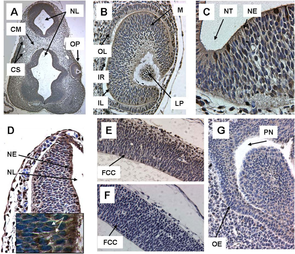
TFII-I expression in head region at E9 (A-D) and at E12 (E, F, G).
Panel
- A. Head region (5 ×) At low power, TFII-I immunoreactivity is visible in multiple regions of the embryo, but is most intense in the neural tube.
- B. Optic vesicle (32 ×) The developing eye demonstrates TFII-I expression in the inner and outer retinal layer as well as lens primordium.
- C. Neural tube (64 ×) Neural tube demonstrates intense nuclear immunoreactivity in individual cells. At E12 TFII-I is expressed extensively in brain, in contrast to TFII-IRD1, whereas TFII-I is only moderately present in nasopharynx.
- D. Brain region (40 ×) Neural tube demonstrates intense nuclear immunoreactivity in individual cells. At E12 TFII-I is expressed extensively in brain, in contrast to TFII-IRD1, whereas TFII-I is only moderately present in nasopharynx.
- E Brain Region (32 ×) TFII-I is expressed in both nuclear and cytoplasmic localization in brain
- F. Brain Region (32 ×) TFII-IRD1 antibody demonstrates no immunoreactivity in an adjacent section.
- G. Nasopharynx (40 ×). Nasopharynx also demonstrates predominantly cytoplasmic localization
Inset in Figure 2D shows nuclear and cytoplasmic locatization of TFII-I under 100 × magnification (Zeiss Axioscop, obj. Apochromat 100 ×). White arrows indicate positive reaction of specific antibody with TFII-I.
Abbr.: CM-Cephalic Mesenchyme, CS-Cardiovascular System, FCC - Future Cerebral Cortex, IL-Inner Retinal Layer, IR-Intra-Retinal Space, LP-Lens Primordium, M-Mesenchyme, NE-Neuroepithelium, NL-Neural Lumen, NT-Neural Tube, OE-Olfactory Epithelium, OL-Outer Retinal Layer, OP-Optic Pit, PN- Primitive Nasopharynx,
Antibodies to the N-terminus of TFII-I were used to probe embryonic mouse sections. TFII-I protein is widely expressed in the developing embryo. TFII-I is expressed throughout the period from E8-E16. However, within this period there are striking shifts in localization from cytoplasmic predominant to nuclear. TFII-I expression varies in both a spatial and temporal fashion. There is extensive expression in neural precursors at E8. This expression persists at later stages. TFII-I is expressed in developing lung, heart and gut structures. There is no evidence of isoform specific expression. Available data regarding expression patterns at both an RNA and protein level throughout development are also comprehensively reviewed.
Figure 2. 1756-0500-3-203-2-l.jpg
Reference
<pubmed>20642858</pubmed>| BMC Res Notes
© 2010 Danoff et al; licensee BioMed Central Ltd. This is an open access article distributed under the terms of the Creative Commons Attribution License (http://creativecommons.org/licenses/by/2.0), which permits unrestricted use, distribution, and reproduction in any medium, provided the original work is properly cited.
File history
Click on a date/time to view the file as it appeared at that time.
| Date/Time | Thumbnail | Dimensions | User | Comment | |
|---|---|---|---|---|---|
| current | 11:58, 8 July 2011 | | 1,000 × 862 (298 KB) | S8600021 (talk | contribs) | TFII-I expression in head region at E9 (A-D) and at E12 (E, F, G). A. Head region (5 ×), B. Optic vesicle (32 ×), C. Neural tube (64 ×), D. Brain region (40 ×), E-F. Brain Region (32 ×), G. Nasopharynx (40 ×). TFII-I is more discretely localized at |
You cannot overwrite this file.
File usage
The following page uses this file: