File:Mouse germinal vesicle 03.jpg

Original file (1,195 × 1,200 pixels, file size: 219 KB, MIME type: image/jpeg)
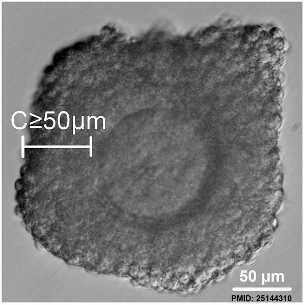
GV stage oocyte classification based on thickness of the cumulus layers
GV stage COCs were collected from ovaries from mice following administration of PMSG. The COCs were classified into three categories according to the thickness of their cumulus layers
(C): COCs with C≥50 µm.
For the experiment using COCs with dispelled cumulus layers, COCs with C≥50 µm (bottom line, left) were treated using gentle pipetting to generate C<30 µm (bottom line, right). Scale = 50 µm.
- Mouse Germinal Vesicle Links: Image - 1 | Image - 2 | Image - 3 | Image - 4 | Oocyte Development | Zona pellucida | Granulosa cell | Mouse Development
Reference
<pubmed>25144310</pubmed>| PLoS One.
Copyright
© 2014 Zhou et al. This is an open-access article distributed under the terms of the Creative Commons Attribution License, which permits unrestricted use, distribution, and reproduction in any medium, provided the original author and source are credited.
File history
Click on a date/time to view the file as it appeared at that time.
| Date/Time | Thumbnail | Dimensions | User | Comment | |
|---|---|---|---|---|---|
| current | 21:36, 8 September 2014 | | 1,195 × 1,200 (219 KB) | Z8600021 (talk | contribs) | ==GV stage oocyte classification based on thickness of the cumulus layers== GV stage COCs were collected from ovaries from mice following administration of PMSG. The COCs were classified into three categories according to the thickness of their cumulu... |
You cannot overwrite this file.
File usage
The following 3 pages use this file: