File:Mouse cornea P0.jpg
Mouse_cornea_P0.jpg (703 × 561 pixels, file size: 101 KB, MIME type: image/jpeg)
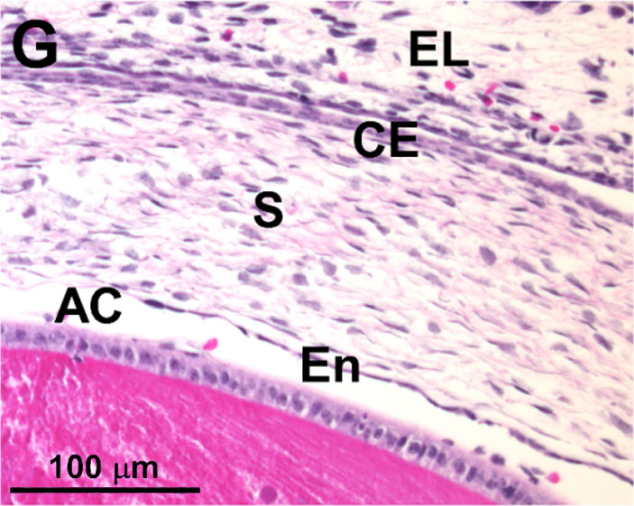
Mouse Corneal Development P0
(Stain - Haematoxylin Eosin)
P0 - Postnatal day 0
corneal stroma (S) Anterior chamber (AC). eyelid (EL) corneal endothelial (En) layer. lens (L) and the corneal epithelium (CE)
- Cornea Links: Image - Mouse cornea development | Image - Mouse E12.5 | Image - Mouse E13.5 | Image - Mouse cell proliferation E13.5 | Image - Mouse E16.5 | Image - Mouse P0 | Cornea Development | Mouse Development
Reference
Zhang J, Upadhya D, Lu L, Reneker LW (2015) Fibroblast Growth Factor Receptor 2 (FGFR2) Is Required for Corneal Epithelial Cell Proliferation and Differentiation during Embryonic Development. PLoS ONE 10(1): e0117089. doi:10.1371/journal.pone.0117089
http://journals.plos.org/plosone/article?id=10.1371/journal.pone.0117089
Copyright
© 2015 Zhang et al. This is an open access article distributed under the terms of the Creative Commons Attribution License, which permits unrestricted use, distribution, and reproduction in any medium, provided the original author and source are credited.
doi:10.1371/journal.pone.0117089.g001
Journal.pone.0117089.g001.jpg Original figure cropped and resized.
File history
Click on a date/time to view the file as it appeared at that time.
| Date/Time | Thumbnail | Dimensions | User | Comment | |
|---|---|---|---|---|---|
| current | 11:50, 24 January 2015 |  | 703 × 561 (101 KB) | Z8600021 (talk | contribs) | ==Mouse Corneal Development== {{HE}} * At E12.5, ocular mesenchymal cells migrated into the space between the lens (L) and the corneal epithelium (CE, arrow in enlarged inset) in eyes. * At E13.5, the corneal epithelial layer * At E16.5, corneal str... |
You cannot overwrite this file.
File usage
The following page uses this file:
