File:Mouse Schlemm's canal development 01.jpg

Original file (1,200 × 873 pixels, file size: 161 KB, MIME type: image/jpeg)
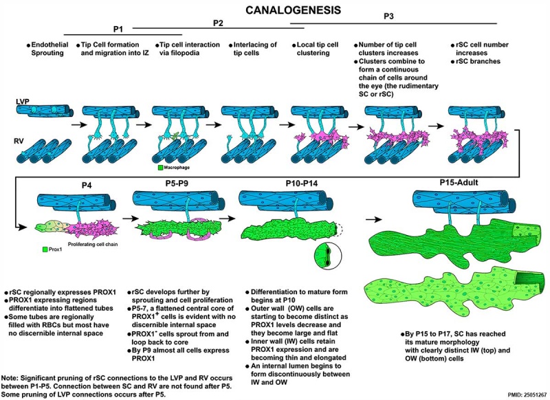
Mouse Schlemm's canal development
Schematic showing the stages of SC development by the novel process of canalogenesis.
Cartoons have been drawn for clarity and are not intended to suggest that most early sprouts arise from the LVP.
Reference
<pubmed>25051267</pubmed>| PLoS Biol.
Copyright
© 2014 Kizhatil et al. This is an open-access article distributed under the terms of the Creative Commons Attribution License, which permits unrestricted use, distribution, and reproduction in any medium, provided the original author and source are credited.
Figure 18. doi:10.1371/journal.pbio.1001912.g018 Original figure cropped, altered in size colour and labelling.
File history
Click on a date/time to view the file as it appeared at that time.
| Date/Time | Thumbnail | Dimensions | User | Comment | |
|---|---|---|---|---|---|
| current | 18:42, 28 January 2015 | | 1,200 × 873 (161 KB) | Z8600021 (talk | contribs) | ==Mouse Schlemm's canal development == Schematic showing the stages of SC development by the novel process of canalogenesis. Cartoons have been drawn for clarity and are not intended to suggest that most early sprouts arise from the LVP. Figure 18.... |
You cannot overwrite this file.
File usage
The following page uses this file: