File:Mouse CT E18.5.jpg
Mouse_CT_E18.5.jpg (221 × 344 pixels, file size: 13 KB, MIME type: image/jpeg)
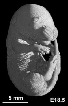
Mouse Embryo
Mouse microCT (E18.5 CRL 16.5mm)
From Fig. 2. Midsagittal slices from volume datasets and surface views of the developing mouse at selected stages (E10.5–PND8).
Reference
<pubmed>18713865</pubmed>| PMID: 18713865 | PNAS
© 2008 by The National Academy of Sciences of the USA
Anyone may, without requesting permission, use original figures or tables published in PNAS for noncommercial and educational use (i.e., in a review article, in a book that is not for sale) provided that the original source and the applicable copyright notice are cited.
File history
Click on a date/time to view the file as it appeared at that time.
| Date/Time | Thumbnail | Dimensions | User | Comment | |
|---|---|---|---|---|---|
| current | 02:29, 16 April 2010 | | 221 × 344 (13 KB) | S8600021 (talk | contribs) | Mouse microCT (E18.5 CRL 16.5mm) From Fig. 2. Midsagittal slices from volume datasets and surface views of the developing mouse at selected stages (E10.5–PND8). High-resolution magnetic resonance histology of the embryonic and neonatal mouse: a 4D atl |
You cannot overwrite this file.
File usage
The following 2 pages use this file: