File:Mouse 3D Heart internal E8.5-14.5.jpeg
From Embryology

Size of this preview: 800 × 374 pixels. Other resolution: 1,280 × 599 pixels.
Original file (1,280 × 599 pixels, file size: 106 KB, MIME type: image/jpeg)
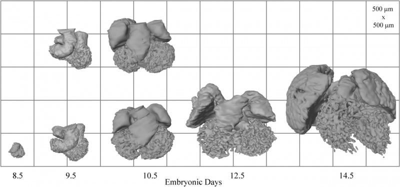
Reconstructions of the lumen of the hearts shown in external view. Duplicate reconstructions are shown for ED 9.5 and 10.5. This series clearly shows the formation of atrial (smooth walled) and ventricular (trabeculated) lumen from the outer curvature of the heart tube.
h71230363005.jpeg
| Species | Stage | |||||||||||||||
| Human | Days | 20 | 22 | 24 | 28 | 30 | 33 | 36 | 40 | 42 | 44 | 48 | 52 | 54 | 55 | 58 |
| Mouse | Days | 9 | 9.5 | 10 | 10.5 | 11 | 11.5 | 12 | 12.5 | 13 | 13.5 | 14 | 14.5 | 15 | 15.5 | 16 |
Reference
Image (used with permission) from the paper. <pubmed>12746463</pubmed>
File history
Click on a date/time to view the file as it appeared at that time.
| Date/Time | Thumbnail | Dimensions | User | Comment | |
|---|---|---|---|---|---|
| current | 20:51, 16 August 2009 |  | 1,280 × 599 (106 KB) | S8600021 (talk | contribs) | Reconstructions of the lumen of the hearts shown in external view. Duplicate reconstructions are shown for ED 9.5 and 10.5. This series clearly shows the formation of atrial (smooth walled) and ventricular (trabeculated) lumen from the outer curvature of |
You cannot overwrite this file.
File usage
The following 3 pages use this file: