File:Mouse-solitary chemosensory cells.jpg

Original file (1,000 × 670 pixels, file size: 89 KB, MIME type: image/jpeg)
Mouse Solitary Chemosensory Cells
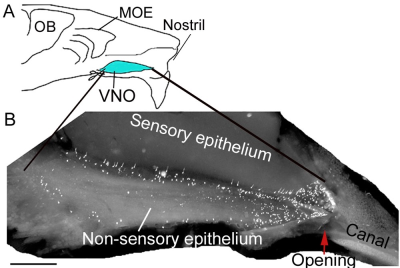
Solitary chemosensory cells (SCCs) preferentially locate at the entrance duct of the VNO and express chemosensory signaling components.
A: A schematic drawing of a mouse hemi-nose. MOE: main olfactory epithelium; OB: olfactory bulb. VNO: vomeronasal organ (blue).
B: Luminal view of the entire non-sensory epithelium and entrance duct of a VNO from a TRPM5-GFP mouse. Bright spots are GFP-positive SCCs. Arrow points to the anterior opening. Anterior to the VNO, the cartilaginous stenonii canal channels external fluids to the VNO opening.
Scale: B, 0.5 mm.
Original File name: Figure 1. http://www.plosone.org/article/slideshow.action?uri=info:doi/10.1371/journal.pone.0011924&imageURI=info:doi/10.1371/journal.pone.0011924.g001
Reference
<pubmed>20689832</pubmed>| PLoS One.
doi:10.1371/journal.pone.0011924.g001
Citation: Ogura T, Krosnowski K, Zhang L, Bekkerman M, Lin W (2010) Chemoreception Regulates Chemical Access to Mouse Vomeronasal Organ: Role of Solitary Chemosensory Cells. PLoS ONE 5(7): e11924. doi:10.1371/journal.pone.0011924
Copyright: © 2010 Ogura et al. This is an open-access article distributed under the terms of the Creative Commons Attribution License, which permits unrestricted use, distribution, and reproduction in any medium, provided the original author and source are credited.
File history
Click on a date/time to view the file as it appeared at that time.
| Date/Time | Thumbnail | Dimensions | User | Comment | |
|---|---|---|---|---|---|
| current | 15:53, 29 September 2010 |  | 1,000 × 670 (89 KB) | S8600021 (talk | contribs) | ==Mouse Solitary Chemosensory Cells== Solitary chemosensory cells (SCCs) preferentially locate at the entrance duct of the VNO and express chemosensory signaling components. A: A schematic drawing of a mouse hemi-nose. MOE: main olfactory epithelium; OB |
You cannot overwrite this file.
File usage
The following page uses this file: