File:Mouse-pituitary Sox4 expression.jpg
Mouse-pituitary_Sox4_expression.jpg (596 × 448 pixels, file size: 55 KB, MIME type: image/jpeg)
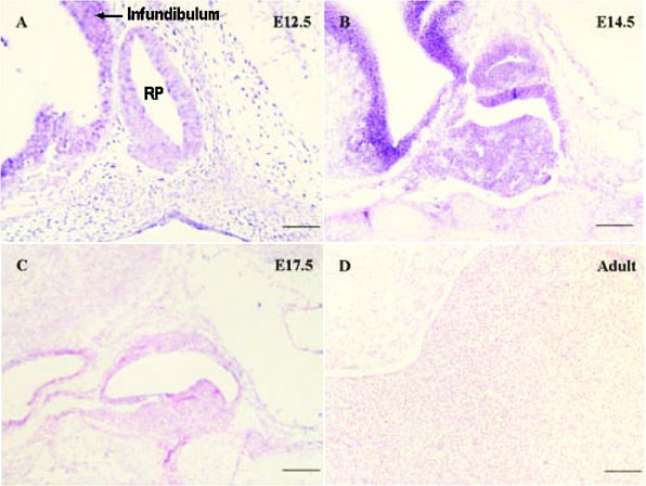
In situ hybridization of Sox4 in mice fetal pituitaries
A-D show the images hybridized with Sox4 probe. The positive blue color was initiated by alkaline phosphatase and BCIP/NBT.
All the slides were counterstained with 0.1% nuclear fast red after in situ hybridization.
Original magnification, ×200 (bar, 50 μm).
- "The significant changes in gene expression in both tissues suggest a distinct and dynamic switch between embryonic and adult pituitaries. All these data along with Sox4 should be confirmed to further understand the community of multiple signaling pathways that act as a cooperative network that regulates maturation of the pituitary. It was also suggested that EST sequencing is an efficient means of gene discovery."
Original file name: Figure 3. 1471-2164-10-109-3.jpg http://www.biomedcentral.com/1471-2164/10/109/figure/F3
Reference
http://www.biomedcentral.com/1471-2164/10/109
Ma et al. BMC Genomics 2009 10:109 doi:10.1186/1471-2164-10-109
© 2009 Ma et al; licensee BioMed Central Ltd.
This is an Open Access article distributed under the terms of the Creative Commons Attribution License (http://creativecommons.org/licenses/by/2.0), which permits unrestricted use, distribution, and reproduction in any medium, provided the original work is properly cited.
File history
Click on a date/time to view the file as it appeared at that time.
| Date/Time | Thumbnail | Dimensions | User | Comment | |
|---|---|---|---|---|---|
| current | 15:37, 2 October 2010 |  | 596 × 448 (55 KB) | S8600021 (talk | contribs) | ==In situ hybridization of Sox4 in mice fetal pituitaries== A-D show the images hybridized with Sox4 probe. The positive blue color was initiated by alkaline phosphatase and BCIP/NBT. All the slides were counterstained with 0.1% nuclear fast red after |
You cannot overwrite this file.
File usage
The following page uses this file:
