File:Mouse-optic nerve axons.jpg

Original file (600 × 693 pixels, file size: 126 KB, MIME type: image/jpeg)
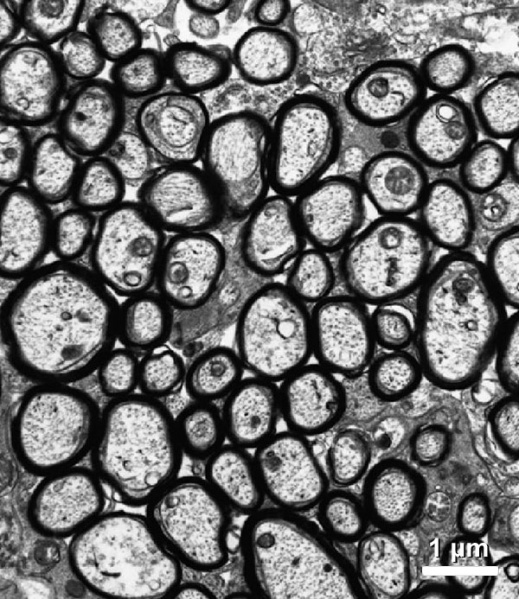
Adult Mouse Optic Nerve Axons
Representative electron micrograph of sections of optic nerve of P28 wild-type mice.
Bar: 1 µm.
Original file name: Figure 4. The relationship between axon diameter and myelin thickness is normal in dnβ1 mice.
image was extracted
Related myelin images: Mouse - sciatic nerve Schwann cell | sciatic nerve | cerebellum axons | optic nerve
Reference
<pubmed>19451276</pubmed>| PMC2711572
This article is distributed under the terms of an Attribution–Noncommercial–Share Alike–No Mirror Sites license for the first six months after the publication date (see http://www.jcb.org/misc/terms.shtml). After six months it is available under a Creative Commons License (Attribution–Noncommercial–Share Alike 3.0 Unported license, as described at http://creativecommons.org/licenses/by-nc-sa/3.0/).
File history
Click on a date/time to view the file as it appeared at that time.
| Date/Time | Thumbnail | Dimensions | User | Comment | |
|---|---|---|---|---|---|
| current | 12:06, 24 September 2010 | | 600 × 693 (126 KB) | S8600021 (talk | contribs) | ==Adult Mouse Optic Nerve Axons== Representative electron micrograph of sections of optic nerve, spinal cord, and cerebellum of P28 wild-type mice. Bar: 1 µm. Original file name: Figure 4. The relationship between axon diameter and myelin thickness is |
You cannot overwrite this file.
File usage
The following 3 pages use this file: