File:Mouse- axial skeleton intervertebral disc.jpg
Mouse-_axial_skeleton_intervertebral_disc.jpg (600 × 300 pixels, file size: 40 KB, MIME type: image/jpeg)
Mouse- axial skeleton intervertebral disc
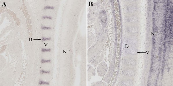
Verification of localization by in situ hybridization.
(A) A digoxigenin labelled probe to Nfatc1 was hybridized to sections from E12.5 day embryos. Hybridization was visualized as purple staining. Nfatc1 was clearly expressed in the IVD (D, arrow) at this stage. (B) A digoxigenin labelled probe to Ebfa1 was hybridized to sections from E12.5 day embryos. Expression was not detected in the IVD
(D) but light purple staining was seen in the vertebral body (V). Staining was also seen in the neural tube (NT) and adjacent blood vessels.
Nfatc1
- Lange AW, Molkentin JD, Yutzey KE: DSCR1 gene expression is dependent on NFATc1 during cardiac valve formation and colocalizes with anomalous organ development in trisomy 16 mice.
Dev Biol 2004, 266:346-60
- Down syndrome critical region 1 (DSCR1) gene encodes a regulatory protein in the calcineurin/NFAT signal transduction pathway (NFATc1)
- Calcineurin signaling through the NFAT family of transcription factors regulates gene expression in response to elevated Ca2+ levels
Ebfa1
- Garel S, Marin F, Mattei MG, Vesque C, Vincent A, Charnay P: Family of Ebf/Olf-1-related genes potentially involved in neuronal differentiation and regional specification in the central nervous system.
Dev Dyn 1997, 210:191-205.
- mouse genes Ebf2 and Ebf3 have high similarity to the rodent Ebf/Olf-1 and the Drosophila collier genes.
- "In the region extending from the midbrain to the spinal cord, the expression of the three genes correlated with neuronal maturation, with a general activation in early post-mitotic cells, followed by specific patterns of extinction also consistent with the neurogenic gradient."
Sohn et al. BMC Developmental Biology 2010 10:29 doi:10.1186/1471-213X-10-29
Original file name: 1471-213X-10-29-2.jpg
Molecular profiling of the developing mouse axial skeleton: a role for Tgfbr2 in the development of the intervertebral disc. Sohn P, Cox M, Chen D, Serra R. BMC Dev Biol. 2010 Mar 9;10:29. PMID: 20214815 | BMC
© 2010 Sohn et al; licensee BioMed Central Ltd.
This is an Open Access article distributed under the terms of the Creative Commons Attribution License (http://creativecommons.org/licenses/by/2.0), which permits unrestricted use, distribution, and reproduction in any medium, provided the original work is properly cited.
File history
Click on a date/time to view the file as it appeared at that time.
| Date/Time | Thumbnail | Dimensions | User | Comment | |
|---|---|---|---|---|---|
| current | 08:32, 28 April 2010 |  | 600 × 300 (40 KB) | S8600021 (talk | contribs) | Mouse- axial skeleton intervertebral disc Verification of localization by in situ hybridization. (A) A digoxigenin labelled probe to Nfatc1 was hybridized to sections from E12.5 day embryos. Hybridization was visualized as purple staining. Nfatc1 was c |
You cannot overwrite this file.
File usage
There are no pages that use this file.
