File:Mouse- adult cortex.jpg
Mouse-_adult_cortex.jpg (464 × 436 pixels, file size: 34 KB, MIME type: image/jpeg)
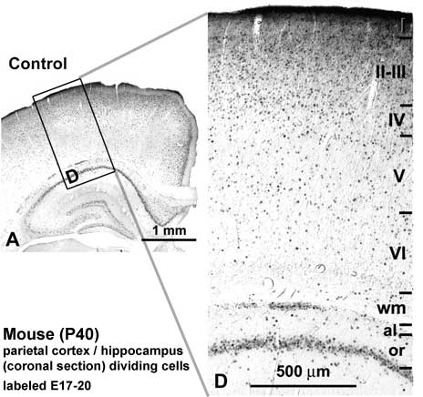
Low-magnification photomicrographs of coronal sections of the parietal cortex and hippocampus showing BrdU-immunoreactive cells after E17–20 injections in control pups at P40.
showing layers I–VI and white matter (wm) the neocortex and the alveus (al) and stratum oriens (or) of the hippocampus.
Original file name: Figure 1. http://www.ncbi.nlm.nih.gov/pmc/articles/PMC2871377/figure/fig1/ (cropped from full figure)
Reference
<pubmed>19812240</pubmed>| PMC2871377
This is an Open Access article distributed under the terms of the Creative Commons Attribution Non-Commercial License (http://creativecommons.org/licenses/by-nc/2.5/uk/) which permits unrestricted non-commercial use, distribution, and reproduction in any medium, provided the original work is properly cited.
File history
Click on a date/time to view the file as it appeared at that time.
| Date/Time | Thumbnail | Dimensions | User | Comment | |
|---|---|---|---|---|---|
| current | 21:56, 14 December 2010 |  | 464 × 436 (34 KB) | S8600021 (talk | contribs) | Low-magnification photomicrographs of coronal sections of the parietal cortex and hippocampus showing BrdU-immunoreactive cells after E17–20 injections in control pups at P40. showing layers I–VI and white matter (wm) the neocortex and the alveus ( |
You cannot overwrite this file.
File usage
The following page uses this file:
