File:Mouse-E12.5.png
Mouse-E12.5.png (496 × 600 pixels, file size: 278 KB, MIME type: image/png)
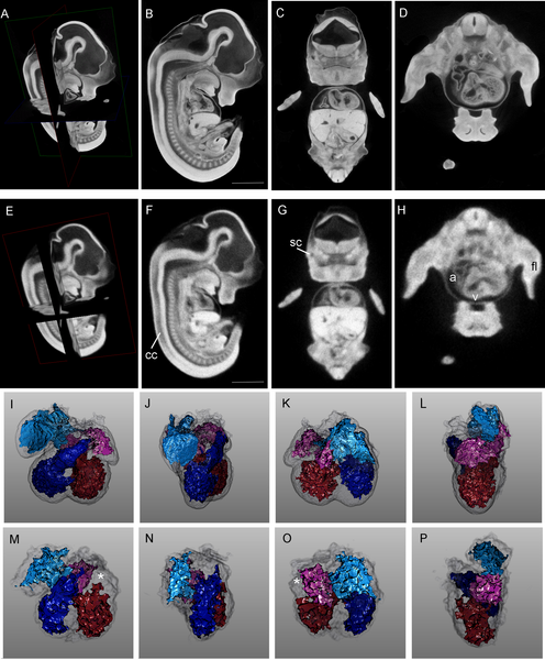
Comparison of High-Resolution (8 μm) and More Rapid (27 μm) Virtual Histology Techniques
(A–D) Wild-type E12.5 embryo scanned at 8-μm resolution. (A) Orientation of the planes of section. (B) Sagittal plane. (C) Coronal plane. (D) Axial plane.
(E–H) The same embryo as in (A–D), scanned by a more rapid protocol at 27-μm resolution. Most of the features present at 8-μm resolution are also appreciable at the lower 27-μm resolution.
(I–P) Comparison of segmentation analysis of cardiac chambers for the 8-μm and 27-μm scans, respectively. The right atrium is teal, the right ventricle is blue, the left atrium is pink, the left ventricle is red, and the cardiac wall is transparent grey. A region of the right atrium that could not be segmented on the 27-μm scan is shown with a white asterisk (*).
Scale bars in panels (B) and (F) represent 1.2 mm. Movies of sagittal, coronal, and axial planes corresponding to panels (B–D) are presented as Videos S1, S2, and S3.
a, cardiac atrium; cc, central canal of the neural tube; fl, forelimb; sc, semicircular canal; v, cardiac ventricle.
Original File Name:Journal.pgen.0020061.g003.png
http://www.plosgenetics.org/article/info:doi/10.1371/journal.pgen.0020061
Citation: Johnson JT, Hansen MS, Wu I, Healy LJ, Johnson CR, et al. (2006) Virtual Histology of Transgenic Mouse Embryos for High-Throughput Phenotyping. PLoS Genet 2(4): e61. doi:10.1371/journal.pgen.0020061
Editor: Wayne Frankel, The Jackson Laboratory, United States of America
Received: January 23, 2006; Accepted: March 13, 2006; Published: April 28, 2006
Copyright: © 2006 Johnson et al. This is an open-access article distributed under the terms of the Creative Commons Attribution License, which permits unrestricted use, distribution, and reproduction in any medium, provided the original author and source are credited.
File history
Click on a date/time to view the file as it appeared at that time.
| Date/Time | Thumbnail | Dimensions | User | Comment | |
|---|---|---|---|---|---|
| current | 00:22, 29 March 2010 |  | 496 × 600 (278 KB) | S8600021 (talk | contribs) | Comparison of High-Resolution (8 μm) and More Rapid (27 μm) Virtual Histology Techniques (A–D) Wild-type E12.5 embryo scanned at 8-μm resolution. (A) Orientation of the planes of section. (B) Sagittal plane. (C) Coronal plane. (D) Axial plane. (E� |
You cannot overwrite this file.
File usage
There are no pages that use this file.
