File:Morphological differences in early mouse embryonic development.png

Original file (2,061 × 886 pixels, file size: 455 KB, MIME type: image/png)
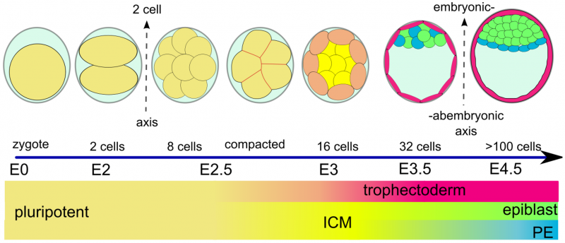
Schematic view of morphological and lineage specification steps during the early mouse embryonic development
Starting from fertilization (E0), after three rounds of cleavages (E1–E2.5), the blastomers undergo compaction and polarization (E3). Then the trophectoderm outer layer starts to separate from the inner cell mass (ICM) followed by the expansion of the blastocoel and the localization of the ICM to one part of the embryo (E3.5). After this stage the endoderm is formed as a layer separating epiblast from blastocoel (E4.5). After 4.5 embryonic days, the preimplantation embryo contains more than 100 cells. http://dx.doi.org/10.1371/journal.pcbi.1001128.g001
Reference
<pubmed>PMC3088645</pubmed> Copyright
© 2011 Krupinski et al. This is an open-access article distributed under the terms of the Creative Commons Attribution License, which permits unrestricted use, distribution, and reproduction in any medium, provided the original author and source are credited.
- Note - This image was originally uploaded as part of an undergraduate science student project and may contain inaccuracies in either description or acknowledgements. Students have been advised in writing concerning the reuse of content and may accidentally have misunderstood the original terms of use. If image reuse on this non-commercial educational site infringes your existing copyright, please contact the site editor for immediate removal.
File history
Click on a date/time to view the file as it appeared at that time.
| Date/Time | Thumbnail | Dimensions | User | Comment | |
|---|---|---|---|---|---|
| current | 12:35, 18 August 2016 | | 2,061 × 886 (455 KB) | Z5019880 (talk | contribs) | PMC3088645 |
You cannot overwrite this file.
File usage
The following page uses this file: