File:Mixed melasma.jpg
From Embryology
Size of this preview: 448 × 600 pixels. Other resolution: 600 × 803 pixels.
Original file (600 × 803 pixels, file size: 112 KB, MIME type: image/jpeg)
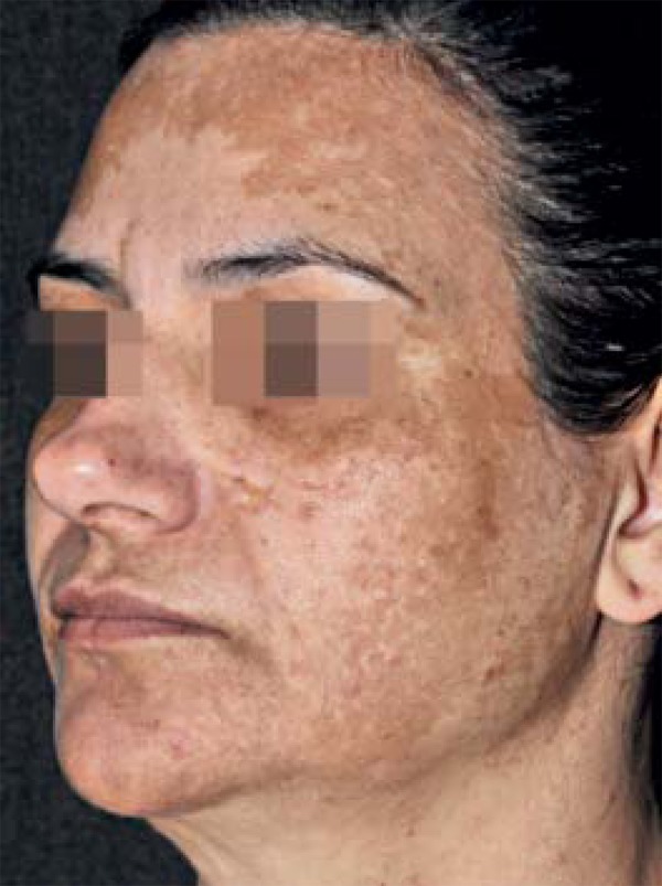
Figure 17: Mixed Facial Melasma
The image shows the mixed facial melasma of a female patient. The melasma is described as mixed as it is a mixture of the identified facial patterns, affecting the frontal, temporal, parotid, mandibular and zygomatic regions.
Reference
Handel AC, Miot LD & Miot HA. (2014). Melasma: a clinical and epidemiological review. An Bras Dermatol , 89, 771-82. PMID: 25184917
Copyright
©2014 by Anais Brasileiros de Dermatologia
- Note - This image was originally uploaded as part of an undergraduate science student project and may contain inaccuracies in either description or acknowledgements. Students have been advised in writing concerning the reuse of content and may accidentally have misunderstood the original terms of use. If image reuse on this non-commercial educational site infringes your existing copyright, please contact the site editor for immediate removal.
File history
Click on a date/time to view the file as it appeared at that time.
| Date/Time | Thumbnail | Dimensions | User | Comment | |
|---|---|---|---|---|---|
| current | 22:45, 16 October 2018 | | 600 × 803 (112 KB) | Z5164785 (talk | contribs) | Figure 17: Mixed Facial Melasma The image shows the mixed facial melasma of a female patient. The melasma is described as mixed as it is a mixture of the identified facial patterns, affecting the frontal, temporal, parotid, mandibular and zygomatic re... |
| 22:30, 16 October 2018 |  | 600 × 803 (112 KB) | Z5164785 (talk | contribs) | Figure 17: Mixed Facial Melasma The image shows the mixed facial melasma of a female patient. The melasma is described as mixed as it is a mixture of the identified facial patterns, affecting the frontal, temporal, parotid, mandibular and zygomatic re... |
You cannot overwrite this file.
File usage
The following 2 pages use this file: