File:Mitochondria EM01.jpg
Mitochondria_EM01.jpg (640 × 480 pixels, file size: 96 KB, MIME type: image/jpeg)
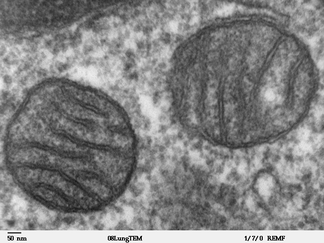
Mitochondria Electron Micrograph
This high magnification image shows two mitochondria within the cytoplasm of a cell, a single cell may contain several 100 mitochondrion. These organelles are the physical location where cell respiration occurs,
Each mitochondrion has the same structure:
- an outer double membrane
- an inter-membranous space
- the inner membrane folded into extensions called cristae
- an internal space called the matrix
Image - Transmission electron microscope photo of a thin section cut through an area of mammalian lung tissue.
- Links: Mitochondria | Category:Mitochondria
Image Source: Contributed by Dartmouth College Electron Microscope Facility special thanks to Chuck Daghlian and Louisa Howard. Gallery. Original images may have been altered in size contrast and labelling. (These images are in the public domain)
JEOL 100CX TEM Writer: Louisa Howard Keywords: mitochondria, mammalian, TEM, lung
Cite this page: Hill, M.A. (2024, April 24) Embryology Mitochondria EM01.jpg. Retrieved from https://embryology.med.unsw.edu.au/embryology/index.php/File:Mitochondria_EM01.jpg
- © Dr Mark Hill 2024, UNSW Embryology ISBN: 978 0 7334 2609 4 - UNSW CRICOS Provider Code No. 00098G
File history
Click on a date/time to view the file as it appeared at that time.
| Date/Time | Thumbnail | Dimensions | User | Comment | |
|---|---|---|---|---|---|
| current | 10:47, 23 June 2011 | | 640 × 480 (96 KB) | S8600021 (talk | contribs) | ==Mitochondria Electron Micrograph== Transmission electron microscope image of a thin section cut through an area of mammalian lung tissue. The high magnification image shows a mitochondria. JEOL 100CX TEM Writer: Louisa Howard Keywords: mitochondria, |
You cannot overwrite this file.
File usage
The following 3 pages use this file: