File:Minot1889 plate27.jpg

Original file (2,000 × 1,496 pixels, file size: 690 KB, MIME type: image/jpeg)
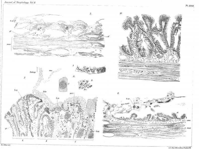
Plate 27.
EXPLANATION OF PLATE XXVII.
Fig. 5. Portion of the ob-placenta of Fig. 2 (x 175 diams.), to show the degenerated epithelium, h.ep, and the saucer-shaped glands, gl, gV.
Fig. 6. Rabbit's uterus of eleven days; portion of the ob-placenta to show the degenerated epithelium, h.ep, and the regenerated glands,^/ ( X 175 diams.).
Fig. 7. Portion of the placenta of Fig. 2 ( X 175 diams.), to show the degeneration of the uterine tissue and the relations of the foetal ectoderm to the placental surface.
Fig. 10. Rabbit's uterus of thirteen days; portion of the ob-placenta (X 175 diams.), to show the regenerated glands.
Fig. II. Portions of the epithelium of the periplacenta at thirteen days. A, vertical section (X 175 diams.). B, surface view (X 175 diams.). C, single cell ( X 445 diams.).
Reference
Minot CS. Uterus And Embryo - I. Rabbit II. Man. (1889) J Morphol. 2:
Cite this page: Hill, M.A. (2024, April 25) Embryology Minot1889 plate27.jpg. Retrieved from https://embryology.med.unsw.edu.au/embryology/index.php/File:Minot1889_plate27.jpg
- © Dr Mark Hill 2024, UNSW Embryology ISBN: 978 0 7334 2609 4 - UNSW CRICOS Provider Code No. 00098G
File history
Click on a date/time to view the file as it appeared at that time.
| Date/Time | Thumbnail | Dimensions | User | Comment | |
|---|---|---|---|---|---|
| current | 13:53, 4 April 2014 | | 2,000 × 1,496 (690 KB) | Z8600021 (talk | contribs) |
You cannot overwrite this file.
File usage
The following page uses this file: