File:Minot1889 plate26.jpg

Original file (2,000 × 1,466 pixels, file size: 587 KB, MIME type: image/jpeg)
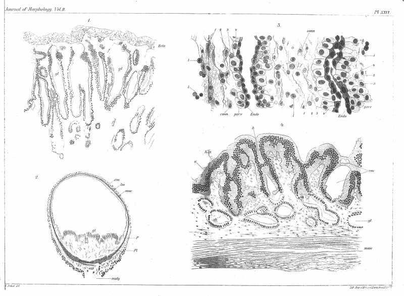
Plate 26.
Fig. I. Placenta of rabbit at eight days, with dilated glands,^/, and superjacent foetal ectoderm, ecto (X 125 diams.).
Fig. 2. Rabbit's uterus at nine days, transverse section of a swelling (X 7 diams.).
Fig. 3. Portion of the placenta of Fig. 2 (X 445 diams.), to show the connective tissue, conn, the perivascular cells, per.v, and the thickened endothelium, endo, of the blood capillaries.
Fig. 4. Portion of the periplacenta of Fig. 2 (X 175 diams.), to show the degeneration of the epithelium, h.ep.
Reference
Minot CS. Uterus And Embryo - I. Rabbit II. Man. (1889) J Morphol. 2:
Cite this page: Hill, M.A. (2024, April 23) Embryology Minot1889 plate26.jpg. Retrieved from https://embryology.med.unsw.edu.au/embryology/index.php/File:Minot1889_plate26.jpg
- © Dr Mark Hill 2024, UNSW Embryology ISBN: 978 0 7334 2609 4 - UNSW CRICOS Provider Code No. 00098G
File history
Click on a date/time to view the file as it appeared at that time.
| Date/Time | Thumbnail | Dimensions | User | Comment | |
|---|---|---|---|---|---|
| current | 13:18, 4 April 2014 | | 2,000 × 1,466 (587 KB) | Z8600021 (talk | contribs) | |
| 13:07, 4 April 2014 |  | 2,000 × 1,466 (623 KB) | Z8600021 (talk | contribs) | {{Minot1889 figures}} |
You cannot overwrite this file.
File usage
The following page uses this file: