File:Minot1889 fig04.jpg
From Embryology

Size of this preview: 800 × 386 pixels. Other resolution: 1,000 × 482 pixels.
Original file (1,000 × 482 pixels, file size: 153 KB, MIME type: image/jpeg)
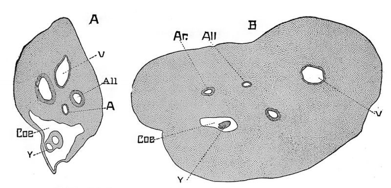
Cut 4. Two sections of umbilical cord
A, at sixty days;
B, at three months;
V, vein; Ar, artery; All, allantois cavity; Coe, coelom; Y, yolk sack ; X 22 diams.
Reference
Minot CS. Uterus And Embryo - I. Rabbit II. Man. (1889) J Morphol. 2:
Cite this page: Hill, M.A. (2024, April 19) Embryology Minot1889 fig04.jpg. Retrieved from https://embryology.med.unsw.edu.au/embryology/index.php/File:Minot1889_fig04.jpg
- © Dr Mark Hill 2024, UNSW Embryology ISBN: 978 0 7334 2609 4 - UNSW CRICOS Provider Code No. 00098G
File history
Click on a date/time to view the file as it appeared at that time.
| Date/Time | Thumbnail | Dimensions | User | Comment | |
|---|---|---|---|---|---|
| current | 12:51, 4 April 2014 | | 1,000 × 482 (153 KB) | Z8600021 (talk | contribs) | no legend |
| 12:50, 4 April 2014 |  | 1,200 × 658 (232 KB) | Z8600021 (talk | contribs) | {{Minot1889 figures}} |
You cannot overwrite this file.
File usage
The following page uses this file: