File:Miller1920 Plate 2.jpg

Original file (987 × 1,200 pixels, file size: 102 KB, MIME type: image/jpeg)
Plate 2
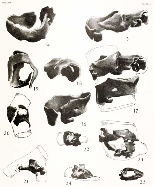
Fig. 14. — Ventro-mesial view of the second right bronchial cartilage. The expanded dorsal end of the mesial arm can be seen; also a portion of the oblong foramen mentioned in the text. X10.
Fig. 15. — Dorsal view of the fused elements that form the anterior cartilage which caps the eparterial bronchus. X10.
Fig. 16. — Dorsal view of the second cartilage associated with the origin of the eparterial bronchus. X10.
Fig. 17. — Dorso-lateral view of the two cartilages which are placed about the origin of the eparterial bronchus from the right bronchus. X 15.
Fig. 18. — Dorso-mesial view of the first cartilage on the bronchus pa.ssing to the left lobus anterior. X15.
Fig. 19. — Ventro-mesial view of the cartilage described in detail in the text. X10.
Fig. 20. — Small bronchus surrounded at its point of origin by three special-shaped cartilages. X10.
Fig. 21. — Dorsal view of a small typical saddle-shaped cartilage shown in situ. Its relation to the two principal bronchi is clearly shown, also the prolongation by which it comes into relation with a third bronchus. The bronchi are shown as though transparent. X10.
Fig. 22. — Top view of a small bronchus surrounded by two saddle-shaped cartilages. X15.
Fig. 23. — A small bronchus surrounded by a ring-shaped cartilage which is quite irregular in its formation. X15.
Fig. 24. — A ring-shaped cartilage which surrounds a small bronchus and by means of a spur partially surrounds a second and smaller bronchus. X15.
Fig. 25. — The cartilage of figure 24 shown independent of its bronchi viewed from above and slightly from ventral side. X10.
Miller Links: Plate 1 | Plate 2 | Contribution No.38 | Volume IX | Contributions to Embryology
| Historic Disclaimer - information about historic embryology pages |
|---|
| Pages where the terms "Historic" (textbooks, papers, people, recommendations) appear on this site, and sections within pages where this disclaimer appears, indicate that the content and scientific understanding are specific to the time of publication. This means that while some scientific descriptions are still accurate, the terminology and interpretation of the developmental mechanisms reflect the understanding at the time of original publication and those of the preceding periods, these terms, interpretations and recommendations may not reflect our current scientific understanding. (More? Embryology History | Historic Embryology Papers) |
Glossary Links
- Glossary: A | B | C | D | E | F | G | H | I | J | K | L | M | N | O | P | Q | R | S | T | U | V | W | X | Y | Z | Numbers | Symbols | Term Link
Cite this page: Hill, M.A. (2024, April 23) Embryology Miller1920 Plate 2.jpg. Retrieved from https://embryology.med.unsw.edu.au/embryology/index.php/File:Miller1920_Plate_2.jpg
- © Dr Mark Hill 2024, UNSW Embryology ISBN: 978 0 7334 2609 4 - UNSW CRICOS Provider Code No. 00098G
File history
Click on a date/time to view the file as it appeared at that time.
| Date/Time | Thumbnail | Dimensions | User | Comment | |
|---|---|---|---|---|---|
| current | 15:11, 14 April 2012 |  | 987 × 1,200 (102 KB) | Z8600021 (talk | contribs) | ==Plate 2== {{Miller1920}} |
You cannot overwrite this file.
File usage
The following page uses this file:
