File:Meyer1932history3 fig01.jpg
Original file (661 × 800 pixels, file size: 147 KB, MIME type: image/jpeg)
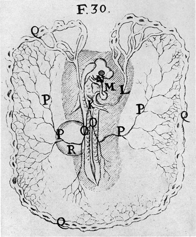
Fig. 1. After Malpighi. Chick after two days’ incubation
K. auricle; L. veins; M, right ventricle; N, left sinus; O, branches of umbilical arteries; P, umbilical arteries; Q, terminations of arteries; R, vesicle near right umbilical branch P is swollen with thin humor or ichor. (This vesicle must have been the extremity of the allantois.)
Reference
Meyer AW. Essays on the History of Embryology: Part III. Cal West Med. 1932 Feb;36(2):105-9. PMID 18742030
Cite this page: Hill, M.A. (2024, April 18) Embryology Meyer1932history3 fig01.jpg. Retrieved from https://embryology.med.unsw.edu.au/embryology/index.php/File:Meyer1932history3_fig01.jpg
- © Dr Mark Hill 2024, UNSW Embryology ISBN: 978 0 7334 2609 4 - UNSW CRICOS Provider Code No. 00098G
File history
Click on a date/time to view the file as it appeared at that time.
| Date/Time | Thumbnail | Dimensions | User | Comment | |
|---|---|---|---|---|---|
| current | 17:07, 2 November 2015 | | 661 × 800 (147 KB) | Z8600021 (talk | contribs) | |
| 17:07, 2 November 2015 | 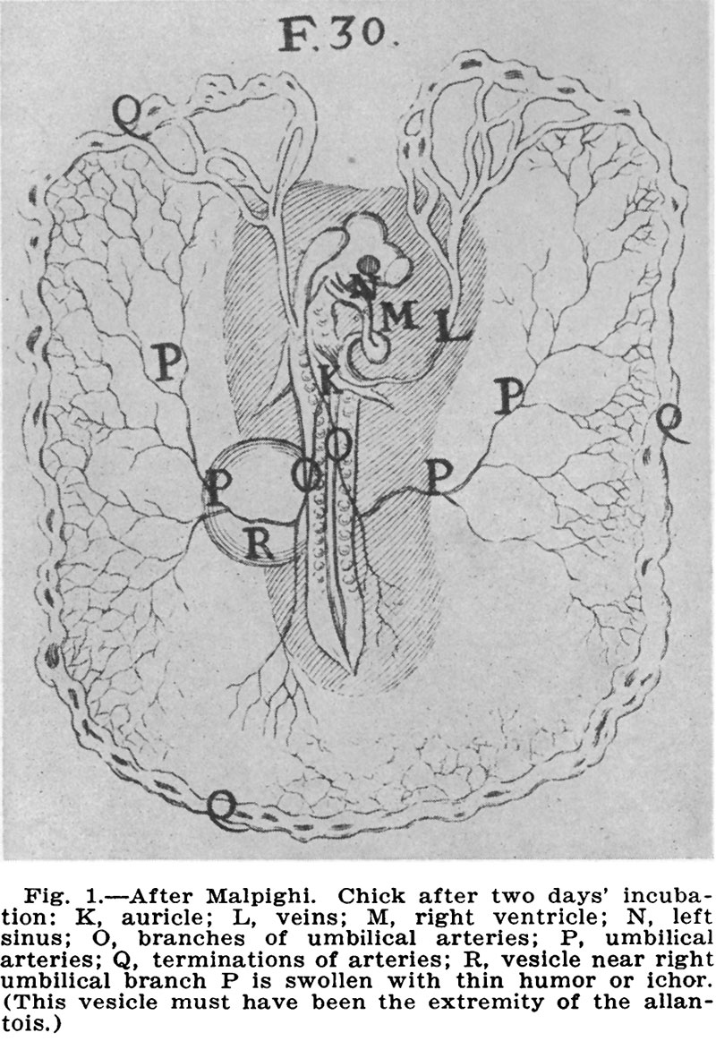 | 800 × 1,163 (215 KB) | Z8600021 (talk | contribs) |
You cannot overwrite this file.
File usage
The following page uses this file: