File:Meyer1920 fig17.jpg
Original file (800 × 703 pixels, file size: 76 KB, MIME type: image/jpeg)
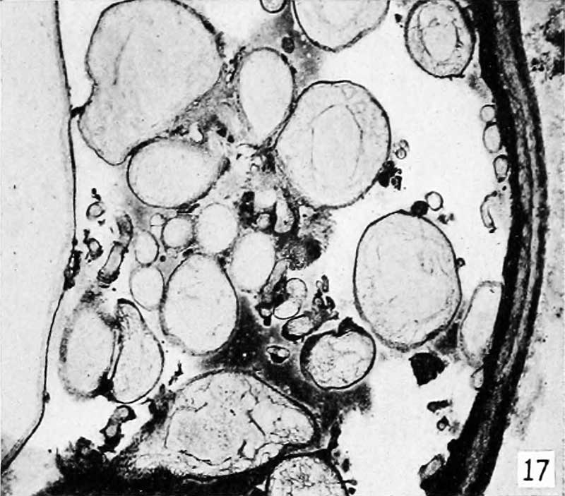
Fig. 17.
It is especially interesting that, just as soon as syncitial hydatitl elliptical villi, or portions of the same begin to appear, the condition can be recognized with some certainty under a magnification of 12 to 20 diameters with the binocular microscope. It often was surprising how relatively early stages could thus be detected and the diagnosis confirmed later by histologic examination. Indeed, Colloidin blocks of tissue from which sections had been cut gave splendid testimony when examined in fluid with the binocular.
One of the not very early stages contained in utero and represented in figure 10 could be recognized with the unaided eye; and when examined with the binocular, under a magnification of about 12 diameters, the picture was unusually fine and wholly unmistakable, as shown in figure 17.
- Meyer Links: Plate 1 | Plate 2 | Plate 3 | Plate 4 | Plate 5 | Plate 6 | Contribution No.40 | Volume IX | Contributions to Embryology | Hydatidiform Mole | Tubal Pregnancy
| Historic Disclaimer - information about historic embryology pages |
|---|
 |
Reference
Meyer AW. Hydatiform degeneration in tubal and uterine pregnancy. (1920) Carnegie Instn. Wash. Publ., Contrib. Embryol., 40: 327- 364.
Cite this page: Hill, M.A. (2024, April 16) Embryology Meyer1920 fig17.jpg. Retrieved from https://embryology.med.unsw.edu.au/embryology/index.php/File:Meyer1920_fig17.jpg
- © Dr Mark Hill 2024, UNSW Embryology ISBN: 978 0 7334 2609 4 - UNSW CRICOS Provider Code No. 00098G
File history
Click on a date/time to view the file as it appeared at that time.
| Date/Time | Thumbnail | Dimensions | User | Comment | |
|---|---|---|---|---|---|
| current | 10:32, 8 April 2012 | | 800 × 703 (76 KB) | Z8600021 (talk | contribs) | ==Fig. 17.== Plate 3: Fig. 14 | Fig. 15 | Fig. 16 | Fig. 17 | Fig. 18 | |
You cannot overwrite this file.
File usage
The following page uses this file: