File:Meyer1920 fig14.jpg
Meyer1920_fig14.jpg (750 × 438 pixels, file size: 31 KB, MIME type: image/jpeg)
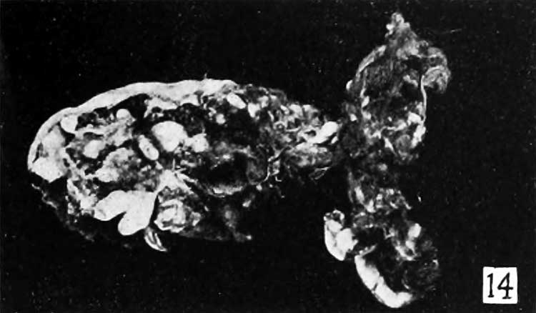
Fig. 14.
No. 1926, a companion specimen to No. 1640, is composed of material from curettage received through the courtesy of Dr. Karl Wilson, of the department of obstetrics of the Johns Hopkins Medical School.
It was removed from the same patient about a year later than specimen No. 1640. Upon gross examination the hydropic nature of some of the villi is plainly evident, as shown in figure 14, and upon microscopic examination the diagnosis of hydatiform degeneration could be confirmed, although the villi were extremely degenerate.
The menstrual history of this case fortunately is known and is thoroughly reliable. The last menstruation occurred January 24 and curettage was done August 4. Bleeding occurred every two or three weeks during March and April and was repeated throughout May. Since the uterus, which had reached the symphysis, had not enlarged any for months, in view of the long duration of pregnancy the operation was performed. The major portion of the specimen is very small. The chorio-decidual portion was felt-like in consistency and extremely fibrous, due largely no doubt to the long retention. Most of the accompanying material looks like mucosa rather than decidua, although some of the larger pieces very evidently contained villi. Some of these were relatively thick and fibrous, and others were vesicular. All of the material was extremely fibrous, making it difficult to get a satisfactory teased preparation.
Accompanying this material was a small body 5X7.5X .30 mm., shown in figure 15. Both nodule and stalk contained some remnants of the embryo. Although the appearance of the stalk suggests the umbilical cord, it contains fragments of the body of the embryo, some of which evidently are composed of nerve tissue.
- Meyer Links: Plate 1 | Plate 2 | Plate 3 | Plate 4 | Plate 5 | Plate 6 | Contribution No.40 | Volume IX | Contributions to Embryology | Hydatidiform Mole | Tubal Pregnancy
| Historic Disclaimer - information about historic embryology pages |
|---|
 |
Reference
Meyer AW. Hydatiform degeneration in tubal and uterine pregnancy. (1920) Carnegie Instn. Wash. Publ., Contrib. Embryol., 40: 327- 364.
Cite this page: Hill, M.A. (2024, April 25) Embryology Meyer1920 fig14.jpg. Retrieved from https://embryology.med.unsw.edu.au/embryology/index.php/File:Meyer1920_fig14.jpg
- © Dr Mark Hill 2024, UNSW Embryology ISBN: 978 0 7334 2609 4 - UNSW CRICOS Provider Code No. 00098G
File history
Click on a date/time to view the file as it appeared at that time.
| Date/Time | Thumbnail | Dimensions | User | Comment | |
|---|---|---|---|---|---|
| current | 10:21, 8 April 2012 | | 750 × 438 (31 KB) | Z8600021 (talk | contribs) | ==Fig. 14.== Plate 3: Fig. 14 | Fig. 15 | Fig. 16 | Fig. 17 | Fig. 18 | |
You cannot overwrite this file.
File usage
The following page uses this file: