File:Meyer1920 fig07.jpg

Original file (482 × 650 pixels, file size: 21 KB, MIME type: image/jpeg)
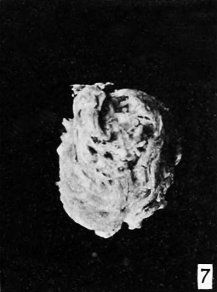
Fig. 7.
A third exceptionally fine specimen of tubal hydatiform mole is No. 2052, donated by Dr. N. M. Davis, of Washington, D. C.
Figure 7 shows a portion of the tube containing the hydatiform mole, some hydatiform villi of which protrude through an incision in the wall of the tube. The whole opening is filled with typical hydatiform villi barely detected by the unaided eye but perfectly evident under an enlargement of 4 diameters. They present an extremely fine picture when seen with the binocular under a magnification of 10 to 20 diameters.
Examination under a higher magnification shows that the preservation of the specimen is unusually good and that all the villi are markedly hydatiform. Trophoblastic proliferation is so marked that in some places it gives the appearance of decidual formation.
Plate 1: Fig. 1 | Fig. 2 | Fig. 3 | Fig. 4 | Fig. 5 | Fig. 6 | Fig. 7
- Meyer Links: Plate 1 | Plate 2 | Plate 3 | Plate 4 | Plate 5 | Plate 6 | Contribution No.40 | Volume IX | Contributions to Embryology | Hydatidiform Mole | Tubal Pregnancy
| Historic Disclaimer - information about historic embryology pages |
|---|
| Pages where the terms "Historic" (textbooks, papers, people, recommendations) appear on this site, and sections within pages where this disclaimer appears, indicate that the content and scientific understanding are specific to the time of publication. This means that while some scientific descriptions are still accurate, the terminology and interpretation of the developmental mechanisms reflect the understanding at the time of original publication and those of the preceding periods, these terms, interpretations and recommendations may not reflect our current scientific understanding. (More? Embryology History | Historic Embryology Papers) |
Reference
Meyer AW. Hydatiform degeneration in tubal and uterine pregnancy. (1920) Carnegie Instn. Wash. Publ., Contrib. Embryol., 40: 327- 364.
Cite this page: Hill, M.A. (2024, April 19) Embryology Meyer1920 fig07.jpg. Retrieved from https://embryology.med.unsw.edu.au/embryology/index.php/File:Meyer1920_fig07.jpg
- © Dr Mark Hill 2024, UNSW Embryology ISBN: 978 0 7334 2609 4 - UNSW CRICOS Provider Code No. 00098G
File history
Click on a date/time to view the file as it appeared at that time.
| Date/Time | Thumbnail | Dimensions | User | Comment | |
|---|---|---|---|---|---|
| current | 09:23, 8 April 2012 |  | 482 × 650 (21 KB) | Z8600021 (talk | contribs) | ==Fig. 1.== Plate 1: Fig. 1 | Fig. 2 | Fig. 3 | Fig. 4 | Fig. 5 | [[:Fi |
You cannot overwrite this file.
File usage
The following 2 pages use this file:
