File:Mesoderm development and Pax 01.jpg

Original file (1,211 × 1,000 pixels, file size: 171 KB, MIME type: image/jpeg)
Mesoderm Development and Pax
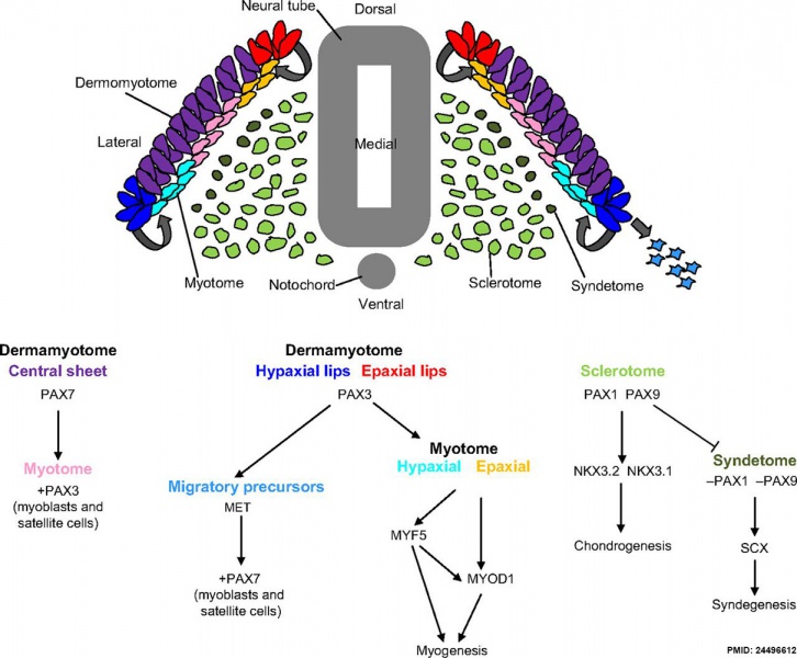
PAX gene hierarchies involved in the development of the progenitor populations of skeletal muscle, cartilage and tendon. Cells in the ventromedial part of the somite de-epithelialise and form the sclerotome (light green), in which Pax1 and Pax9 are expressed in a Shh-dependent manner.
As the dermomyotome elongates dorsomedially and ventrolaterally, PAX3 becomes restricted to the epaxial (red) and hypaxial (dark blue) lips, where a secondary generation of myocytes delaminate and migrate to form the epaxial (orange) and hypaxial (light blue) myotome. In the limb region, PAX3+ progenitors delaminate from the hypaxial dermamyotome (light blue) and migrate into the developing limbs to provide a pool of progenitors. In these cells, PAX3 regulates the hepatocyte growth factor receptor Met, which is necessary for hypaxial delamination and migration.
Pax7 is expressed principally within the central sheet of the dermomyotome (purple); when the final somite dissociates, Pax7+ progenitors delaminate into the developing myotome. These myotomal Pax3+ Pax7+ cells (pink) retain a progenitor state in order to become a resident population necessary for skeletal muscle growth as development proceeds.
The syndetome (dark green) originates from the dorsolateral edge of the sclerotome, as Pax1 and Pax9 are downregulated and scleraxis (Scx) upregulation leads to syndegenesis. Pax, paired homeobox; MYOD1, myogenic differentiation antigen 1; MYF5, myogenic factor 5; NKX, NK homeobox; SCX, scleraxis.
(Text modified from figure legend)
- Links: image - Pax and DNA | image - Pax and Mesoderm | image - somite components | PAX | somitogenesis | skeletal muscle | tendon
Reference
Blake JA & Ziman MR. (2014). Pax genes: regulators of lineage specification and progenitor cell maintenance. Development , 141, 737-51. PMID: 24496612 DOI.
Copyright
This is an Open Access article distributed under the terms of the Creative Commons Attribution License (http://creativecommons.org/licenses/by/3.0), which permits unrestricted use, distribution and reproduction in any medium provided that the original work is properly attributed.
Figure 4 http://dev.biologists.org/content/141/4/737/F4.expansion.html Original figure resized and relabelled. Text above modified from original figure legend.
Cite this page: Hill, M.A. (2024, April 24) Embryology Mesoderm development and Pax 01.jpg. Retrieved from https://embryology.med.unsw.edu.au/embryology/index.php/File:Mesoderm_development_and_Pax_01.jpg
- © Dr Mark Hill 2024, UNSW Embryology ISBN: 978 0 7334 2609 4 - UNSW CRICOS Provider Code No. 00098G
File history
Click on a date/time to view the file as it appeared at that time.
| Date/Time | Thumbnail | Dimensions | User | Comment | |
|---|---|---|---|---|---|
| current | 10:22, 8 July 2014 |  | 1,211 × 1,000 (171 KB) | Z8600021 (talk | contribs) | ==Mesoderm Development and Pax== Pax gene hierarchies involved in the development of the progenitor populations of skeletal muscle, cartilage and tendon. Cells in the ventromedial part of the somite de-epithelialise and form the sclerotome (light gree... |
You cannot overwrite this file.
File usage
The following 3 pages use this file: