File:McMurrich1930 fig87.jpg
Original file (1,280 × 1,612 pixels, file size: 267 KB, MIME type: image/jpeg)
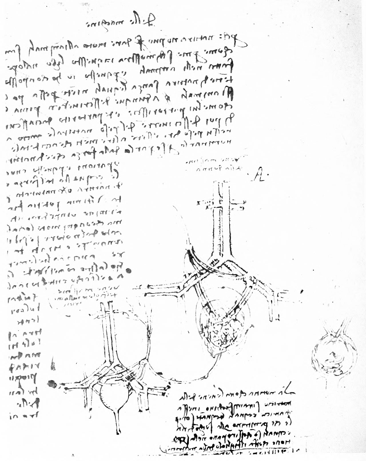
Fig. 87. Diagram of the Human Fetal Circulation
(QI, 1.)
Another figure, on QI, 1, is of interest because it represents the supposed human conditions (fig. 87). The lower portions of the aorta and inferior vena cava and their division into the iliac arteries and veins are shown and below is an oval structure, apparently representing the uterus, within which is a mass which is presumably meant for the discoidal placenta. To this an artery and vein (uterine) pass from each of the iliac vessels— for the vessels passing upward from the iliacs, see p. 176 — and from the placenta there passes a single structure, evidently the umbilical cord. This extends to the fetal umbilicus and there gives off the ascending umbilical vein and the fetal hypogastric arteries. It seems probable that Leonardo believed the hypogastric vessels to carry vital spirit to the fetus, while nourishment was carried by the umbilical vein, since in another passage (QIII, 7v) the blood is said to pass from the fetal liver to the stomach, where it is converted into chyle; this passes to the intestine where a portion is absorbed by the meseraic veins, the rest remaining in the intestine and forming the meconium or fetal faeces.
The structure of the cotyledons interested him greatly. He recognized that at birth each cotyledon divides, part remaining connected with the uterus and part adhering to the chorion (QIII, 8; AnB, 29v), and gives figures (fig. 87) showing the separation (QIII, 8; AnB, 29v, 38), but is somewhat uncertain as to their actual structure.
Reference
McMurrich JP. Leonardo da Vinci - the anatomist. (1930) Carnegie institution of Washington, Williams & Wilkins Company, Baltimore.
Cite this page: Hill, M.A. (2024, April 20) Embryology McMurrich1930 fig87.jpg. Retrieved from https://embryology.med.unsw.edu.au/embryology/index.php/File:McMurrich1930_fig87.jpg
- © Dr Mark Hill 2024, UNSW Embryology ISBN: 978 0 7334 2609 4 - UNSW CRICOS Provider Code No. 00098G
File history
Click on a date/time to view the file as it appeared at that time.
| Date/Time | Thumbnail | Dimensions | User | Comment | |
|---|---|---|---|---|---|
| current | 09:56, 25 March 2020 | | 1,280 × 1,612 (267 KB) | Z8600021 (talk | contribs) | BW and scaled to 1280 pixels wide |
| 09:56, 25 March 2020 |  | 2,224 × 3,266 (392 KB) | Z8600021 (talk | contribs) | Fig. 87. Diagram of the human fetal circulation. (QI, 1.) ===Reference=== {{Ref-McMurrich1930}} {{Footer}} Category:Historic EmbryologyCategory:FetalCategory:Placenta |
You cannot overwrite this file.
File usage
The following 2 pages use this file: