File:MartinFalkiner1938 fig05.jpg
From Embryology

Size of this preview: 410 × 599 pixels. Other resolution: 700 × 1,022 pixels.
Original file (700 × 1,022 pixels, file size: 88 KB, MIME type: image/jpeg)
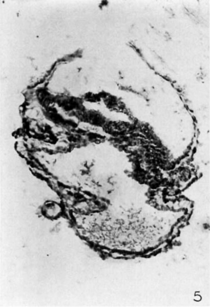
Fig. 5
Section 30 B. X150. This section passes through the primitive streak, which is seen at the caudal half of the embryonic plate. The amnion and embryonie plate have been torn. A t0ng'ue of tissue still pa.1'tia.lly divides the yolk sac.
Reference
Martin CP. and Falkiner N. Mcl. The Falkiner ovum. (1938) Amer. J Anat., 63: 251-271.
Cite this page: Hill, M.A. (2024, April 25) Embryology MartinFalkiner1938 fig05.jpg. Retrieved from https://embryology.med.unsw.edu.au/embryology/index.php/File:MartinFalkiner1938_fig05.jpg
- © Dr Mark Hill 2024, UNSW Embryology ISBN: 978 0 7334 2609 4 - UNSW CRICOS Provider Code No. 00098G
File history
Click on a date/time to view the file as it appeared at that time.
| Date/Time | Thumbnail | Dimensions | User | Comment | |
|---|---|---|---|---|---|
| current | 12:49, 11 August 2017 |  | 700 × 1,022 (88 KB) | Z8600021 (talk | contribs) |
You cannot overwrite this file.
File usage
The following 3 pages use this file: