File:Mall1916 plate03.jpg

Original file (1,316 × 1,500 pixels, file size: 305 KB, MIME type: image/jpeg)
Plate 3.
Fig. 1. Ovum No. 545. X7. There is a delicate network of fibrils below the amnion and the chorion.
Fig. 2. Embryo No. .588. XS. Delicate strands are shown radiating from the umbilical cord and yolk-sac. This figure is given to show the appearance of magma in vesicle development. From a woman who has had numerous mechanical abortions performed upon herself. Uterus badly inflamed.
Fig. 3. Section through the chorion and magma of No. 402. X280. The specimen was stained with Van Gieson stain and shows that tlie fibrils of the magma are continuous witli those of the mesenchyme of the chorionic wall. It came from a case with subinvolution and symptoms of endometritis.
Fig. 4. Outline of the ovum of No. 660. natural size. The diagram indicates the part of the specimen shown enlarged in figure 5.
Fig. 5. No. 660, showing very extensive changes in the magma. X6. The upper tip of the amnion is shown. The magma is fibrillar and granular, and at places the fibrils seem to form membranes. The chorionic wall is very hemorrhagic.
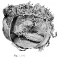
Fig. 1
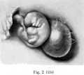
Fig. 2
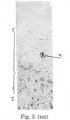
Fig. 3
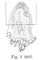
Fig. 4

Fig. 5
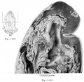
Fig. 4 and Fig 5


| Historic Disclaimer - information about historic embryology pages |
|---|
 |
- Mall 1916 Links: Table 1 Normal Embryos | Table 2 Abnormal Embryos |Plate 1 | Fig. 1+2 | Fig. 1 | Fig. 2 | Fig. 3 | Fig. 4 | Fig. 5 | Fig. 6 | Fig. 7 | Fig. 8 | Fig. 9 | Fig. 10 | Plate 2 | Fig. 1 | Fig. 2 | Fig. 3 | Plate 3 | Fig. 1 | Fig. 2 | Fig. 3 | Fig. 4+5 | Fig. 4 | Fig. 5 | Contributions to Embryology No.10
File history
Click on a date/time to view the file as it appeared at that time.
| Date/Time | Thumbnail | Dimensions | User | Comment | |
|---|---|---|---|---|---|
| current | 11:28, 22 April 2014 | | 1,316 × 1,500 (305 KB) | Z8600021 (talk | contribs) | ==Plate 3. == {{Mall1916 figures}} |
You cannot overwrite this file.
File usage
The following page uses this file: