File:Mall1916 fig18.jpg
From Embryology
Size of this preview: 513 × 599 pixels. Other resolution: 1,294 × 1,511 pixels.
Original file (1,294 × 1,511 pixels, file size: 279 KB, MIME type: image/jpeg)
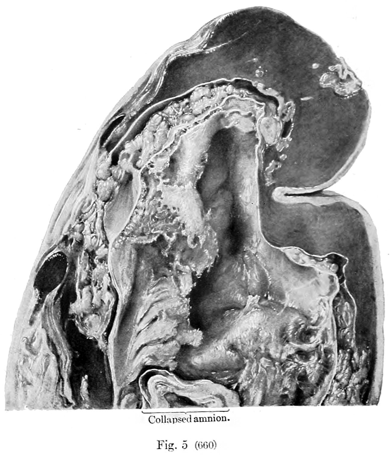
Fig.5. Human Embryo No. 660
No. 660, showing very extensive changes in the magma. X6. The upper tip of the amnion is shown. The magma is fibrillar and granular, and at places the fibrils seem to form membranes. The chorionic wall is very hemorrhagic.
| Historic Disclaimer - information about historic embryology pages |
|---|
 |
- Mall 1916 Links: Table 1 Normal Embryos | Table 2 Abnormal Embryos |Plate 1 | Fig. 1+2 | Fig. 1 | Fig. 2 | Fig. 3 | Fig. 4 | Fig. 5 | Fig. 6 | Fig. 7 | Fig. 8 | Fig. 9 | Fig. 10 | Plate 2 | Fig. 1 | Fig. 2 | Fig. 3 | Plate 3 | Fig. 1 | Fig. 2 | Fig. 3 | Fig. 4+5 | Fig. 4 | Fig. 5 | Contributions to Embryology No.10
File history
Click on a date/time to view the file as it appeared at that time.
| Date/Time | Thumbnail | Dimensions | User | Comment | |
|---|---|---|---|---|---|
| current | 11:29, 22 April 2014 | | 1,294 × 1,511 (279 KB) | Z8600021 (talk | contribs) | {{Mall1916 figures}} |
You cannot overwrite this file.
File usage
The following 2 pages use this file: