File:Mall1908a plate01.jpg
Original file (568 × 900 pixels, file size: 70 KB, MIME type: image/jpeg)
Plate I
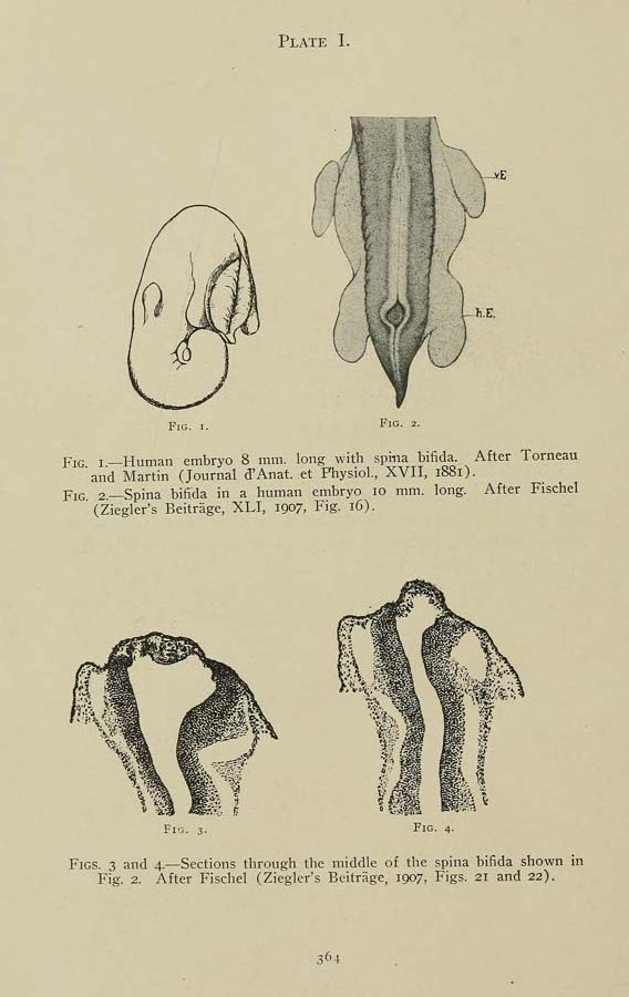
Fig. 1. Human embryo 8 mm. long. with spina bifida. After Tomeau and Martin (Journal d’Anat. et Physiol., XVII, 1881).
Fig. 2. Spina bifida in a human embryo 10 mm. long. After Fischel (Ziegler’s Beitriige, XLI, 1907, Fig. 16).
Fig. 3 and 4. Sections through the middle of the spina bifida shown in Fig. 2. After Fischel (Ziegler’s Bertrfige, 1907, Figs, 21 and 22),
| Historic Disclaimer - information about historic embryology pages |
|---|
 |
Mall 1908: Plates | plate 1 | plate 2 | plate 3 | fig 8 | fig 9 | plate 4 | fig 10 | fig 11 | Figures
Reference
Mall FP. A study of the causes underlying the origin of human monsters. (1908) J Morphol. 19: 3-368.
Cite this page: Hill, M.A. (2024, April 19) Embryology Mall1908a plate01.jpg. Retrieved from https://embryology.med.unsw.edu.au/embryology/index.php/File:Mall1908a_plate01.jpg
- © Dr Mark Hill 2024, UNSW Embryology ISBN: 978 0 7334 2609 4 - UNSW CRICOS Provider Code No. 00098G
File history
Click on a date/time to view the file as it appeared at that time.
| Date/Time | Thumbnail | Dimensions | User | Comment | |
|---|---|---|---|---|---|
| current | 17:23, 25 July 2018 | | 568 × 900 (70 KB) | Z8600021 (talk | contribs) |
You cannot overwrite this file.
File usage
The following page uses this file: