File:MRI placenta uterus upper segment 37 weeks gestation.jpg

Original file (1,200 × 773 pixels, file size: 109 KB, MIME type: image/jpeg)
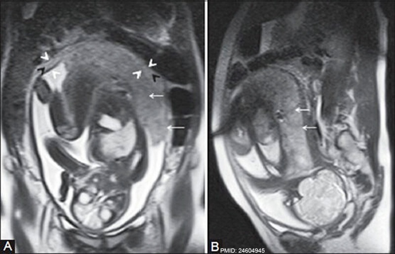
MRI placenta uterus upper segment 37 weeks gestation
Normal mature placenta at upper segment of uterus at 37 weeks of gestation. (A, B) Coronal and sagittal T2 HASTE MR images show a mildly heterogeneous placenta with normal placental septi (white arrows) and triple-layered appearance of normal myometrium (white and black arrowheads)
Reference
<pubmed>24604945</pubmed>PMC3932583 | Indian J Radiol Imaging.
Varghese B, Singh N, George RA, Gilvaz S. Magnetic resonance imaging of placenta accreta. Indian J Radiol Imaging [serial online] 2013 [cited 2014 Mar 18];23:379-85. Available from: http://www.ijri.org/text.asp?2013/23/4/379/125592
Copyright
© 2007 - 2014 Indian Journal of Radiology and Imaging
File history
Click on a date/time to view the file as it appeared at that time.
| Date/Time | Thumbnail | Dimensions | User | Comment | |
|---|---|---|---|---|---|
| current | 15:25, 6 June 2014 |  | 1,200 × 773 (109 KB) | Z8600021 (talk | contribs) | ==MRI placenta uterus upper segment 37 weeks gestation== :'''Links:''' Placenta Development | Magnetic Resonance Imaging ===Reference=== <pubmed>24604945</pubmed>[http://www.ncbi.nlm.nih.gov/pmc/articles/PMC3932583 PMC3932583] | [http://www.i... |
You cannot overwrite this file.
File usage
There are no pages that use this file.